Posterior longitudinal ligament
| Posterior longitudinal ligament | |
|---|---|
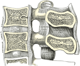
 Posterior longitudinal ligament, in the thoracic region. (Posterior longitudinal ligament runs vertically at center.) | |
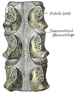
 Median sagittal section of two lumbar vertebrae and their ligaments. (Posterior longitudinal ligament runs vertically at center left.) | |
| Details | |
| Identifiers | |
| Latin | ligamentum longitudinale posterius |
| TA | A03.2.01.008 |
| FMA | 31894 |
| Anatomical terminology | |
The posterior longitudinal ligament is situated within the vertebral canal, and extends along the posterior surfaces of the bodies of the vertebrae, from the body of the axis, where it is continuous with the tectorial membrane of atlanto-axial joint, to the sacrum.[1]
It is broader above than below, and thicker in the thoracic than in the cervical and lumbar regions. The ligament is more narrow at the vertebral bodies and wider at the intervertebral disc space which is more pronounced than the anterior longitudinal ligament. This is significant in understanding certain pathological conditions of the spine such as the typical location for a spinal disc herniation.
In the situation of the intervertebral fibrocartilages and contiguous margins of the vertebrae, where the ligament is more intimately adherent, it is broad, and in the thoracic and lumbar regions presents a series of dentations with intervening concave margins; but it is narrow and thick over the centers of the bodies, from which it is separated by the basivertebral veins.
This ligament is composed of smooth, shining, longitudinal fibers, denser and more compact than those of the anterior ligament, and consists of superficial layers occupying the interval between three or four vertebræ, and deeper layers which extend between adjacent vertebrae.
It functions to prevent hyperflexion of the vertebral column.
See also
References
This article incorporates text in the public domain from page 288 of the 20th edition of Gray's Anatomy (1918)
- ↑ Nigel Palastanga; Roger W. Soames (2012). Churchill Livingstone, ed. Anatomy and Human Movement: Structure and Function.
Additional images
 F: Posterior longitudinal ligament
 Membrana tectoria, transverse, and alar ligaments.
External links
- Atlas image: back_bone25 at the University of Michigan Health System - "Vertebral Column, Dissection, Anterior & Posterior Views"
- lesson7 at The Anatomy Lesson by Wesley Norman (Georgetown University) - "Lateral Pharyngeal Region"