N-localizer
| N-localizer | |
|---|---|
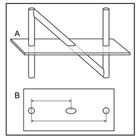 Depiction of the N-localizer and its intersection with the computed tomography (CT) image plane. (A) Side view of the N-localizer. The CT image plane intersects two vertical rods and one diagonal rod. (B) CT image. The intersection of the CT image plane with the N-localizer creates two fiducial circles and one fiducial ellipse. The relative spacing between the ellipse and the two circles varies according to the height at which the CT image plane intersects the diagonal rod. Measuring this spacing permits calculation of the point where the CT image plane intersects the diagonal rod. | |
| Intervention | radiosurgery |
The N-localizer or N-bar is a device that enables guidance of stereotactic surgery or radiosurgery using tomographic images that are obtained via medical imaging technologies such as X-ray computed tomography (CT), magnetic resonance imaging (MRI) or positron emission tomography (PET).[1][2]
Additional images

Figure 2. Three N-localizers are attached to a stereotactic instrument. The three N-localizers are placed around the hemi circumference of the stereotactic instrument. The instrument shown is a prototype that was built in order to test the concept of the N-localizer.[3] The small spheres inside the instrument were used to simulate intracranial tumors. The vertical rod at the right rear of the instrument is larger in diameter than the other rods. This large rod facilitates unambiguous interpretation of the fiducial landmarks in a tomographic image.[4]
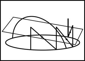
Figure 3. Depiction of three N-localizers and their intersection with the computed tomography (CT) image plane. The quadrilateral represents the CT image plane. The oval and the arch represent the stereotactic instrument. The vertical and diagonal lines that are attached to the oval represent the three N-localizers. The three points where the CT image plane intersects the diagonal rods are depicted by the dots. These points of intersection determine the spatial orientation of the CT image plane relative to the stereotactic instrument, and permit the transfer of patient information from the two-dimensional coordinate system of the planar image into the three-dimensional coordinate system of the stereotactic instrument.
References
- ↑ Galloway, RL Jr. (2015). "Introduction and Historical Perspectives on Image-Guided Surgery". In Golby, AJ. Image-Guided Neurosurgery. Academic Press. pp. 3–4. doi:10.1016/B978-0-12-800870-6.00001-7. ISBN 978-0-12-800870-6.
- ↑ Tse, VCK; Kalani, MYS; Adler, JR (2015). "Techniques of Stereotactic Localization". In Chin, LS; Regine, WF. Principles and Practice of Stereotactic Radiosurgery. New York: Springer. p. 28.
- ↑ Brown, Russell A. (1979). "A computerized tomography-computer graphics approach to stereotaxic localization". Journal of Neurosurgery. 50 (6): 715–20. doi:10.3171/jns.1979.50.6.0715. PMID 374688.
- ↑ Brown RA, Roberts TS, Osborn AG (1981). "Simplified CT-guided stereotaxic biopsy". American Journal of Neuroradiology. 2 (2): 181–184. PMID 6784559.
Further reading
- Brown, Russell A; Nelson, James A (2015). "The Origin and History of the N-Localizer for Stereotactic Neurosurgery". Cureus. doi:10.7759/cureus.323. ISSN 2168-8184.
This article is issued from
Wikipedia.
The text is licensed under Creative Commons - Attribution - Sharealike.
Additional terms may apply for the media files.