Michael I. Miller
| Michael I. Miller | |
|---|---|
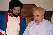 Michael I. Miller (left) and Ulf Grenander in Mittag-Leffler Institute in Stockholm, Sweden circa summer 1995. | |
| Born |
1955 (age 62–63) Brooklyn, New York, United States |
| Residence | United States |
| Nationality | American |
| Alma mater |
The State University of New York at Stony Brook The Johns Hopkins University |
| Known for | Computational anatomy[1] |
| Spouse(s) | Elizabeth Patton Miller[2] |
| Children | 1 |
| Awards |
Presidential Young Investigator Award Johns Hopkins University Gilman Scholar[3] |
| Scientific career | |
| Fields |
Biomedical Engineering Neuroscience Pattern Theory |
| Institutions |
Washington University in St. Louis The Johns Hopkins University |
| Thesis | Statistical Coding of Complex Speech Stimuli in the Auditory Nerve (1983) |
| Doctoral advisor | Murray B. Sachs[4] |
| Website | |
Michael Ira Miller (born 1955) is an American-born biomedical engineer and neuroscientist, and the Massey Professor and Director of the Johns Hopkins University Department of Biomedical Engineering. He worked with Ulf Grenander in the field of Computational Anatomy, specializing in mapping the brain using medical imaging. Miller is the director of the Johns Hopkins Center for Imaging Science, Whiting School of Engineering and codirector of Johns Hopkins Kavli Neuroscience Discovery Institute with Richard L. Huganir. Miller is also a Johns Hopkins University Gilman Scholar.[5]
Biography
In 1976, Miller received his Bachelor of Engineering from The State University of New York at Stony Brook. He then received his Master of Science and PhD in biomedical engineering from the Johns Hopkins University.[6]
He worked at Washington University in St. Louis on medical imaging with Donald L. Snyder, then chair of Electrical Engineering at Washington University School of Engineering and Applied Science. He then joined the faculty of Electrical Engineering in 1985 and remained there through 1998 as the Newton R. and Sarah Louisa Glasgow Wilson Professor in Engineering.[7] From 1994 through 2001, Miller was a visiting professor at Brown University's Division of Applied Mathematics, where he worked with Ulf Grenander on image analysis.
In 1998 Miller joined the Department of Biomedical Engineering at Johns Hopkins University, where he is currently the Massey Professor and Director of Biomedical Engineering, as well as the director of the Center for Imaging Science. In March 2011, Miller was appointed by President Ronald J. Daniels as one of 17 inaugural University Gilman Scholars.[5] In 2015, Miller became the co-director of the newly established Kavli Institute for Discovery Neuroscience.[8] In July, 2017 he became the director of the Department of Biomedical Engineering at the Johns Hopkins University.[6]
Academic career
Neural coding
Miller did his doctoral work on neural codes in the Auditory system under the direction of Murray B. Sachs and Eric D. Young in the Neural Encoding Laboratory[9] at Johns Hopkins University. With Sachs and Young, Miller focused on rate-timing population codes of complex features of speech including voice-pitch[10] and consonant-vowel syllables [11] encoded in the discharge patterns across the primary auditory nerve. These neural codes were one of the scientific works discussed as a possible basis for the discussions at the 1982 New York Academy of Science[12] meeting on the efficacy and timeliness of Cochlear implants.
Medical imaging
Miller's work in the field of brain mapping via Medical imaging, specifically statistical methods for iterative image reconstruction, began in the mid 1980s when he joined Donald L. Snyder at Washington University to work on time-of-flight positron emission tomography (PET) systems being instrumented in Michel Ter-Pogossian's group. With Snyder, Miller worked to stabilize likelihood-estimators of radioactive tracer intensities via the method-of-sieves[13] .[14] This became one of the main approaches for controlling noise artifacts in the Shepp-Vardi algorithm[15] in the context of low-count, time-of-flight emission tomography. It was during this period that Miller met Lawrence (Larry) Shepp, and he subsequently visited Shepp several times at Bell Labs to speak as part of the Henry Landau seminar series.
Pattern theory and computational anatomy
During the mid 1990s, Miller joined the Pattern Theory group at Brown University and worked with Ulf Grenander on problems in image analysis within the Bayesian framework of Markov random fields. They established the ergodic properties of jump-diffusion processes for inference in hybrid parameter spaces, which was presented by Miller at the Journal of the Royal Statistical Society as a discussed paper. [16] These were the first results on a class of random sampling algorithms with ergodic properties proven to sample from distributions supported across discrete sample spaces and simultaneously over the continuum, likening it to the extremely popular Gibb's sampler of Geman and Geman[17] as well as more classical diffusion-based samplers associated to Langevin dynamics.
Grenander and Miller introduced Computational anatomy as a formal theory of human shape and form at a joint lecture in May 1997 at the 50th Anniversary of the Division of Applied Mathematics at Brown University,[18] and in a subsequent publication.[19] In the same year with Paul Dupuis, they published the foundational paper establishing the necessary Sobolev smoothness conditions requiring vector fields to have strictly greater than 2.5 square-integrable, generalized derivatives (in space of 3-dimensions) to ensure that smooth submanifold shapes are carried smoothly by the flows.[20] By 2005, the Computational anatomy framework establishing high-dimensional brain mapping via diffeomorphisms at the morphological scale of MRI had become the de facto standard for cross-section analyses of populations studied via 1mm MRI. Codes now exist for diffeomorphic template or atlas mapping, including ANTS,[21] DARTEL,[22] DEMONS,[23] LDDMM,[24] StationaryLDDMM,[25] all actively used codes for constructing correspondences between coordinate systems based on sparse features and dense images.
Shape and form
David Mumford appreciated the smoothness results on existence of flows, and encouraged collaboration between Miller and the École normale supérieure de Cachan group that had been working independently. In 1998, Mumford organized a Trimestre on "Questions Mathématiques en Traitement du Signal et de l'Image" at the Institute Henri Poincaré; from this emerged the ongoing collaboration on shape between Miller, Alain Trouve and Laurent Younes.[26] They published three significant papers together over the subsequent 15 years; the equations for geodesics generalizing the Euler equation on fluids supporting localized scale or compressibility appeared in 2002,[27] the conservation of momentum law for shape momentum appeared in 2006,[28] and the summary of Hamiltonian formalism appeared in 2015.[29]
Neurodegeneration in brain mapping
Miller and John Csernansky developed a long-term research effort on neuroanatomical phenotyping of Alzheimer's disease, Schizophrenia and mood disorder. In 2005, they published with John Morris an early work on predicting conversion to Alzheimer's disease based on clinically available MRI measurements using diffeomorphometry technologies.[30] This was one of the papers that contributed to a deeper understanding of the disorder in its earlier stages and the recommendations of the working group to revise the diagnostic criteria for Alzheimer’s disease dementia for the first time in 27 years.[31]
In 2009, the Johns Hopkins University BIOCARD[32] project was initiated, led by Marilyn Albert, to study preclinical Alzheimer's disease. In 2014, Miller and Younes demonstrated that the original Braak staging of the earliest change associated to the entorhinal cortex in the medial temporal lobe could be demonstrated via diffeomorphometry methods in the population of clinical MRIs,[33] and subsequently that this could be measured via MRI in clinical populations upwards of 10 years before clinical symptoms appeared.[34]
Books
References
- ↑ Grenander, Ulf; Miller, Michael I. (December 1998). "Computational Anatomy: An Emerging Discipline". Quarterly of Applied Mathematics. 56 (4): 617–694. JSTOR 43638257.
- ↑ Patton Miller, Elizabeth. "Johns Hopkins Humanities Center".
- ↑ "University taps 17 as inaugural Gilman Scholars". The JHU Gazette. Johns Hopkins. 2011.
- ↑ Sachs, M.B. (February 2002). "Member of National Academy of Engineering".
- 1 2 "University taps 17 as inaugural Gilman Scholars". The JHU Gazette. Johns Hopkins. 14 March 2011.
- 1 2 "Michael Miller Named Director of Biomedical Engineering - 06/30/2017". Retrieved 2017-12-09.
- ↑ "Newton R. and Sarah Louisa Glasgow Wilson Professorship in Engineering" (PDF).
- ↑ "RESEARCH: New Kavli Neuroscience Discovery Institute at The Johns Hopkins University | Johns Hopkins Whiting School of Engineering". Johns Hopkins Whiting School of Engineering. 2015-10-02. Retrieved 2017-12-09.
- ↑ "Neural Encoding Laboratory".
- ↑ Miller, M.I.; Sachs, M.B. (June 1984). "Representation of voice pitch in discharge patterns of auditory-nerve fibers". Hearing Research. 14 (3): 257–279. doi:10.1016/0378-5955(84)90054-6. PMID 6480513.
- ↑ Miller, M.I.; Sachs, M.B. (1983). "Representation of stop consonants in the discharge patterns of auditory-nerve fibers". JASA. 74 (2): 502–517. doi:10.1121/1.389816.
- ↑ Sachs, M.B.; Young, E.D.; Miller, M.I. (June 1983). "Speech Encoding in the Auditory Nerve: Implications for Cochlear Implants". Annals of the New York Academy of Sciences. 405: 94–114. doi:10.1111/j.1749-6632.1983.tb31622.x.
- ↑ Snyder, Donald L.; Miller, Michael I. (1985). "The Use of Sieves to Stabilize Images Produced with the EM Algorithm for Emission Tomography". IEEE Transactions on Nuclear Science. NS-32(5): 3864–3872. doi:10.1109/TNS.1985.4334521.
- ↑ Snyder, D.L.; Miller, M.I.; Thomas, L.J.; Politte, D.G. (1987). "Noise and edge artifacts in maximum-likelihood reconstructions for emission tomography". IEEE Trans. on Medical Imaging. 6 (3): 228–238. doi:10.1109/tmi.1987.4307831.
- ↑ Shepp, L.; Vardi, Y. (1982). "Maximum likelihood reconstruction for emission tomography". IEEE Transactions Medical Imaging. 1 (2): 113–122. doi:10.1109/TMI.1982.4307558. PMID 18238264.
- ↑ Grenander, U.; Miller, M.I. (1994). "Representations of Knowledge in Complex Systems". Journal of the Royal Statistical Society, Series B. 56 (4): 549–603. JSTOR 2346184.
- ↑ S. Geman; D. Geman (1984). "Stochastic Relaxation, Gibbs Distributions, and the Bayesian Restoration of Images". IEEE Transactions on Pattern Analysis and Machine Intelligence. 6 (6): 721–741. doi:10.1109/TPAMI.1984.4767596.
- ↑ Walter Freiberger (ed.). "Current and Future Challenges in the Applications of Mathematics". Quarterly of Applied Mathematics.
- ↑ Grenander, Ulf; Miller, M.I. (December 1998). "Computational Anatomy: An Emerging Discipline" (PDF). Quarterly of Applied Mathematics. LVI (4): 617–694.
- ↑ Dupuis, P.; Grenander, U.; Miller, M.I. (September 1998). "Variational Problems on Flows of Diffeomorphisms for Image Matching". Quarterly of Applied Mathematics. 56 (3): 587–600. JSTOR 43638248.
- ↑ "stnava/ANTs". GitHub. Retrieved 2015-12-11.
- ↑ Ashburner, John (2007-10-15). "A fast diffeomorphic image registration algorithm". NeuroImage. 38 (1): 95–113. doi:10.1016/j.neuroimage.2007.07.007. PMID 17761438.
- ↑ "Software - Tom Vercauteren". sites.google.com. Retrieved 2015-12-11.
- ↑ "NITRC: LDDMM: Tool/Resource Info". www.nitrc.org. Retrieved 2015-12-11.
- ↑ "Publication:Comparing algorithms for diffeomorphic registration: Stationary LDDMM and Diffeomorphic Demons". www.openaire.eu. Retrieved 2015-12-11.
- ↑ DOUCET, Sandra. "CMLA - CMLA Research Center for Applied Maths". cmla.ens-paris-saclay.fr. Retrieved 2017-12-06.
- ↑ Miller, M.I.; Trouve, A.; Younes, L. (2002). "On the Metrics and Euler-Lagrange Equations of Computational Anatomy". Annual Review of Biomedical Engineering. 4: 375–405. doi:10.1146/annurev.bioeng.4.092101.125733. PMID 12117763.
- ↑ Miller, M.I.; Trouve, A.; Younes, L. (31 January 2006). "Geodesic shooting for computational anatomy". International Journal of Computer Vision. 24 (2): 209–228. doi:10.1007/s10851-005-3624-0. PMC 2897162. PMID 20613972.
- ↑ Miller, M.I.; Trouve, A.; Younes, L. (December 2015). "Hamiltonian Systems and Optimal Control in Computational Anatomy: 100 Years Since D'Arcy Thompson". Annual Review of Biomedical Engineering. 17: 447–509. doi:10.1146/annurev-bioeng-071114-040601. PMID 26643025.
- ↑ Csernansky, J.G.; Wang, L.; Swank, J.; Miller, JP; Gado, M.; McKeel, D.; Miller, M.I.; Morris, J.C. (15 April 2005). "Preclinical detection of Alzheimer's disease: hippocampal shape and volume predict dementia onset in the elderly". NeuroImage. 25 (3): 783–792. doi:10.1016/j.neuroimage.2004.12.036. PMID 15808979.
- ↑ "Alzheimer's Diagnostic Guidelines". Division of Neuroscience.
- ↑ Albert, M. S. "BIOCARD: Predictors of Cognitive Decline Among Normal Individuals". Alzheimer's Disease Research Center. Johns Hopkins University School of Medicine.
- ↑ Miller, M.I.; Younes, L.; Ratnanather, J.T.; Brown, T.; Trinh, H.; Postal, E.; Lee, D.S.; Wang, M.C; Mori, S.; Obrien, R.; Albert, M.; Research Team, BIOCARD (16 September 2013). "The diffeomorphometry of temporal lobe structures in preclinical Alzheimer's disease". NeuroImage: Clinical. 3 (352–360): 352–360. doi:10.1016/j.nicl.2013.09.001.
- ↑ Younes, L.; Albert, M.; Miller, M.I.; Research Team, BIOCARD (21 April 2014). "Inferring changepoint times of medial temporal lobe morphometric change in preclinical Alzheimer's disease". NeuroImage: Clinical. 5: 178–187. doi:10.1016/j.nicl.2014.04.009. PMC 4110355. PMID 25101236.
External links
| Wikimedia Commons has media related to Michael I. Miller. |
| Authority control |
|---|