Max Brödel
| Max Brödel | |
|---|---|
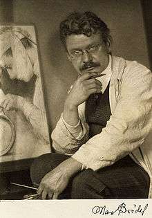 Photograph of Max Brödel by Doris Ulmann | |
| Born |
8 June 1870 Leipzig, Germany |
| Died | 26 October 1941 (aged 71) |
| Nationality | German |
| Education | Leipzig Academy of Fine Arts |
| Known for | Medical Illustration |
| Spouse(s) | Ruth Huntington |
Max Brödel (June 8, 1870 – October 26, 1941) was a medical illustrator. Born in Leipzig, Germany, he began his artistic career after graduating from the Leipzig Academy of Fine Arts, working for Dr. Carl Ludwig. Under Dr. Carl Ludwig's instruction, he gained a basic knowledge of medicine and became recognized for his detailed medical illustrations. In the late 1890s, he was brought to the Johns Hopkins School of Medicine in Baltimore to illustrate for Harvey Cushing, William Halsted, Howard Kelly, and other notable clinicians. In addition to being an prolific medical illustrator, he developed new artistic techniques such as the carbon dust technique that helped the advancement of the quality and accuracy of medical illustrations for physicians. In 1911, he presided over the creation of the first Department of Art as Applied to Medicine; located at the Johns Hopkins School of Medicine, it continues to train medical illustrators to this day. His graduates spread out across the world, and have founded a number of other academic programs.
Biography
Early life and education
Max Brödel was born on June 8, 1870 in Leipzig, Germany, to Louis Brödel and Henrietta Frenzel Brödel. From the early age of 6, he took piano lessons and by 12, he was playing Beethoven. Not only was he musically inclined, he was also artistically inclined. At age 15, Brödel began to develop his artistic abilities at the Leipzig Academy of Fine Arts. The artistic techniques he learned there reflected the 19th century arts education emphasis on the development of fine, precise drawings.[1] This meticulous attention to detail and accuracy was one of the skills that Brödel was later praised for in his medical illustrations. Over the summers, he put his artistic skills to use with part-time jobs drawing landscapes and figures. Brödel was 18 when he had his first experience with medical illustrations, which he would make his lifelong career.
Personal life
Marriage and family
Max Brödel was introduced to fellow artist, medical illustrator, and future wife, Ruth Huntington, by Dr. Howard Kelly. An artist as well, Ruth also received Franklin P. Malls' invitation and had begun illustrating for Dr. Charles Bardeen as part of the Hopkins Anatomy Department in 1900.[2] The pair realized their similar musical and artistic interests and married shortly afterwards on December 31, 1902. They had four children together: Elizabeth (born October 9, 1903), Ruth (born April 23, 1905), Carl (born June 7, 1908), and Elsa (born February 8, 1911). Ruth suffered from scarlet fever as a child and died on June 1, 1908.[3] Elizabeth later followed her father's foosteps and became a medical illustrator.
He was known for his jovial, fun-loving personality, he was a close friend of H.L. Mencken and regular at the Saturday Night Club, where he frequented to drink and enjoy himself.[4] In his free time, he enjoyed fishing and playing the piano.[5] Outside of his profession, he also occasionally made drawings from nature.[2]
Early career
Despite his minimal scientific background and lack of medical knowledge, Brödel and his artistic potential were well received by esteemed German physician and physiologist, Carl Ludwig. Under Ludwig’s mentorship and guidance at the Anatomical Institute at the Institute of Physiology at the University of Leipzig, Brödel was employed with drawing detailed gross anatomical and histological diagrams. Honing his observational skills with detailed notes of the numerous surgeries and autopsies he observed, Brödel’s work was credited for topographical accuracy, tissue realism, and attention to the cross-sectional anatomy.[1] Another noticeable feature of his illustrations was the aerial perspective that showed the anatomy as seen through a surgeon's eyes.[6] Some of his early illustrations were also for physicians Spalteholz, His and Braune.[6] His network of medical professionals increased when he met Dr. Franklin P. Mall of Johns Hopkins Hospital in 1888.[2]
Brödel's artistic career was briefly suspended when he was drafted to serve two years on November 8, 1890.[2] Through the auspices of Geheimrat Carl Ludwig, Prince George of Saxony, Brödel served his first year with arms, and the second year with artistic pursuits for the regiment. Upon return to Leipzig after his service, Brödel continued his work as a free-lance artist, specializing in anatomical and scientific illustrations. During this time, Brödel accepted Mall's invitation to illustrate at Johns Hopkins Hospital.[2]
Career at Johns Hopkins University
Brödel arrived at Johns Hopkins in the winter of January 18, 1894. Highly sought after by anatomist Franklin P. Mall and other physicians for his meticulous attention to detail and realism in his medical illustrations, Brödel's skills were a valuable asset to the Johns Hopkins Hospital. Shortly after his employment, Brödel was joined by fellow medical illustrators, Hermann Becker and August Horn, both of whom had also attended the Leipzig Academy of Fine Arts. Working in conjunction with these two artists, Brödel created an extensive catalog of gross and histological diagrams for the medical staff.[2]
Work with Dr. Howard Kelly
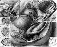
The majority of Brödel's illustrations were for Dr. Howard A. Kelly (1858-1943), the Chief of Gynecology, during his employment at Johns Hopkins Hospital. Brödel illustrated for Kelly's two-volume textbook, Operative Gynecology, which was published in 1898. Its release garnered widespread praise and recognition, cemented Kelly's preeminent status in the field of gynecology, and established Brödel's role as a pioneering medical illustrator. Brödel then went on to work on other books authored or co-authored by Kelly, including those on diseases of the kidneys, ureters and bladder, as well as Kelly’s journal articles and monographs.[3] Throughout the illustrative process, Brödel worked closely with Kelly, conferring with each other before the first sketch was drawn. After debriefing, with Kelly, Brödel painstakingly conducted independent medical research and experimented to find the best method to communicate information about complex structures to medical professionals.
Brödel's underlying artistic philosophy is best described in his own words: “The artist must first fully comprehend the subject matter from every standpoint: anatomical, topographical, histological, pathological, medical, and surgical. From this accumulated knowledge grows a mental picture from which again crystallizes the plan for the future drawing. A clear and vivid mental picture must always precede the actual picture on paper.The planning of the picture, therefore, is the all important thing, not the execution.”[2] His emphasis on anatomically accurate visualization prior to artistic actualization was manifested in his incredibly lifelike renderings.
The seamless translation of medical knowledge into his illustrations is credited with his strong investigative drive. Brödel understood the essential role medical illustrations played in teaching medical students the complexities and functions of anatomical structures, and was therefore keen on educating himself by poring over medical texts, attending lectures, and disseecting cadavers.[7]
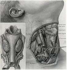
Other medical fields he worked extensively in are Otolaryngology and Urology.[7]
Setbacks
On March 24, 1899, Max Brödel was diagnosed with a streptococcus infection on his hand and arm, caused by improper practice of handling anatomical dissections without gloves.[2] He required several operations on his left arm, including one to separate nerve fibers from the scar tissue. These operations were performed by Dr. William S. Halsted, Chief of Surgery at Johns Hopkins Hospital. Capitalizing on this experience, Brödel illustrated and detailed his medical condition and the resulting numbness of his nondominant left hand. Despite encouragement by Halsted, these drawings remained unpublished.[2]
In December 1904, Brödel sustained severe injuries to the middle finger of his right hand. Another Johns Hopkins physician, Dr. John Miller Turpin Finney, was able to help recover normal functioning, allowing Brödel to continue his artistic and musical pursuits.[2]
War years
With the onset of World War I, Brödel experienced alienation and disillusion living amongst anti-German sentiment in the United States along with his mother’s declining health back in Germany. Henriette Brödel would end up dying November 2, 1915 and Max would become more introverted as the years went on, realizing he had overestimated the amount of importance and growth his medical illustration training program was to receive, expecting it to grow in stature in ways it never did.
Max’s program was to be plagued by low student enrollment during the war years and the persistent troubles of meager compensation in the profession of medical illustration, with two of his pupils turning down offers to work with Brödel’s former colleague Harvey Cushing, now at Harvard Medical School, over the issue of salary.[3]
Famous works and techniques
Carbon dust technique
Max Brödel is credited with the development of the carbon dust technique for medical and scientific illustrations. He had been looking for an acceptable medium able to show the vividness and detail characteristic of living tissue, and made the breakthrough using clay-surfaced lithographic transfer paper.[3] Using a wide variety of media, realistic multi-dimensional representations of complex anatomical structures are able to be constructed. The dust is made by shaving carbon pencils against abrasive surfaces, and then applying this fine dust onto textured, calcium-coated paper with dry brushes. Increasing the depth and dimension of the image, the carbon dust technique was able to add highlights, shadows, and texture to Brödel's work. Due to the limitations of the black and white printing era, the relative ease of reprinting artwork created with carbon dust made this a highly suitable technique for a wide variety of scientific illustrations.[8] Popularized in the 1900s, this method is still used today because of its ability to capture a remarkable amount of fine visual detail.
Department of Art as Applied to Medicine
In 1910, Brödel received an inviting offer for a position at the Mayo Clinic. Gynecologist and close friend of Brödel, Dr. Thomas S. Cullen, began raising funds for a department where Brödel could remain content at Johns Hopkins and train the next generation of medical illustrators with the necessary skills and background.[3]
Henry Walters, a Baltimore financier, philanthropist and art collector, agreed to fund the creation of this endeavor.[9] In 1911, Brödel became the inaugural director for the Department of Art as Applied to Medicine at Johns Hopkins. His goal was to train medical illustrators to work in conjunction with physicians to increase understanding of how the body works. The program was the first of its kind, and attracted both medical and art students from all around the world.
In an article published in the September 1911 edition of The Johns Hopkins Hospital Bulletin, Brödel laid out his case for the creation of the department. “Its purpose,” he wrote, “is to bridge over the gap existing between art and medicine, and to train a new generation of artists to illustrate medical journals and books in the future and
to spare them the years of trial and disappointment of their self-taught predecessors.”[10]
Brödel headed the department until 1939.[1]
Death
Max Brödel died on October 26, 1941 of pancreatic cancer in Baltimore, Maryland.[7] Approximately two months before he passed away, Brödel published a paper in the Journal of the American Medical Association titled "Medical Illustration." This provided a first-hand account and insight into his long illustrative career.[2]
Illustrative legacy
Notable textbooks
- Operative Gynecology (Vols. I&II), Kelly
- Gynecology, Kelly
- Medical Gynecology, Kelly
- The Vermiform Appendix and Its Diseases, Kelly, and Elizabeth Herndon
- Gynecology and Abdominal Surgery (Vols. I&II), Kelly and Charles Noble
- Myomata of the Uterus, Kelly and Cullen
- Diseases of the Kidneys, Ureters and Bladder (Vols. I&II), Kelly and Charles Burnham
Department of Art as Applied to Medicine
Brödel not only leaves behind a massive collection of medical illustrations, his carbon dust technique is still employed today, and the Department of Art as Applied to Medicine is still recognized for their excellence in visual communication in science and medicine. Many former students at the Department of Art as Applied to Medicine would later make up a large percentage of the founding members of the Association of Medical Illustrators, which began in 1945.[3] Several notable artists who were heavily influenced by Brödel include the following.[3]
- Elizabeth Brödel of the Woman's Clinic in the New York Hospital
- James F. Didusch of the Department of Embryology in the Carnegie Institution
- Dorcas Hager Padget of Johns Hopkins School of Medicine
- Willard C. Shepard of Chicago
- Leon Schlossberg of Johns Hopkins School of Medicine
Institutions that have been influenced by Brödel’s work in medical illustrations include the Wilmer, Brady, Mayo and Lahey clinics, the American Museum of Natural History, and Yale, Minnesota, Rochester, Toronto and Tulane Universities.[6]
Johns Hopkins Hospital
In 1938, a portrait of Max Brödel by artist Thomas C. Corner, was presented and displayed in the halls of the Johns Hopkins University School of Medicine alongside portraits of medical pioneers, William Osler, Wiliam Stewart Halsted, Howard Atwood Kelly, and William H. Welch.[6] This display of recognition was initiated by the vice president of the W.B. Saunders medical publishing company, Mr. R.W. Greene.
Brödel Archives
The majority of Brödel's illustrations and his uncompleted manuscript are housed in the Brödel archives located at the Johns Hopkins School of Medicine.[1] Visitors and researchers are allowed to reproduce a selection of his works with special permission. All of Brödel's work for Dr. Kelly and Dr. Thomas S. Cullen are numbered from 1 to 989.
See also
References
- 1 2 3 4 Medicine, Faculty of. "Max Brodel (1870-1941): His Artistic Influence on Surgical Learning at Johns Hopkins Medical School". www.med.uottawa.ca. Retrieved 2017-04-23.
- 1 2 3 4 5 6 7 8 9 10 11 Cullen, Thomas S. (2017-04-23). "Max Brödel, 1870-1941 Director of the First Department of Art as Applied to Medicine in the World". Bulletin of the Medical Library Association. 33 (1): 4.1–29. ISSN 0025-7338. PMC 200894.
- 1 2 3 4 5 6 7 Crosby, Ranice (1991). Max Brödel: The Man Who Put Art Into Medicine. Springer. ISBN 0-387-97563-2.
- ↑ "Max Brodel". Johns Hopkins Magazine. Retrieved 2017-03-27.
- ↑ Cullen, Thomas (1945). Max Brödel, 1870-1941, Director of the First Department of Art as Applied to Medicine in the World. Bulletin of the Medical Library Association. PMC 200894.
- 1 2 3 4 "MAX BRODEL AND MEDICAL ILLUSTRATION". Journal of the American Medical Association. 110 (11). 1938-03-12. doi:10.1001/jama.1938.02790110043013. ISSN 0002-9955.
- 1 2 3 Medicine, Faculty of. "Max Brodel (1870-1941): His Artistic Influence on Surgical Learning at Johns Hopkins Medical School". www.med.uottawa.ca. Retrieved 2017-04-23.
- ↑ 1937-2006., Hodges, Elaine R. S.,; (U.S.), Guild of Natural Science Illustrators (1989-01-01). The Guild handbook of scientific illustration. Wiley. ISBN 9780471288961. OCLC 41168198.
- ↑ Melloni, Ida Dox (1990). Max Brödel and visual communication: The effect of the Hopkins intellectual context in the genesis of modern medical illustration. University of Maryland College Park: UMI.
- ↑ Johns Hopkins Hospital. Bulletin of the Johns Hopkins Hospital. Baltimore: The Hospital, 1891.
Further reading
- Ranice W. Crosby; John Cody (1991). Max Brödel: The Man Who Put Art Into Medicine. Berlin: Springer. ISBN 0-387-97563-2.
External links
- Department of Art as Applied to Medicine
- Association of Medical Illustrators
- Works by or about Max Brödel at Internet Archive
| Authority control |
|---|