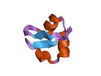
In molecular biology the LysM domain is a protein domain found in a wide variety of extracellular proteins and receptors. The LysM domain is named after the Lysin Motif which was the original name given to the sequence motif identified in bacterial proteins. The region was originally identified as a C-terminal repeat found in the Enterococcus hirae muramidase.[1] The LysM domain is found in a wide range of microbial extracellular proteins, where the LysM domain is thought to provide an anchoring to extracellular polysaccharides such as peptidoglycan and chitin. LysM domains are found in plant receptor kinases for the Nod factor where they bind to symbiotic bacteria in the root nodule. The LysM domain is typically between 44 and 65 amino acid residues in length.[2] The structure of the LysM domain showed that it is composed of a pair of antiparallel beta strands separated by a pair of short alpha helices.[3]
References
- ↑ Joris B, Englebert S, Chu CP, Kariyama R, Daneo-Moore L, Shockman GD, Ghuysen JM (March 1992). "Modular design of the Enterococcus hirae muramidase-2 and Streptococcus faecalis autolysin". FEMS Microbiology Letters. 70 (3): 257–64. PMID 1352512.
- ↑ Buist G, Steen A, Kok J, Kuipers OP (May 2008). "LysM, a widely distributed protein motif for binding to (peptido)glycans". Molecular Microbiology. 68 (4): 838–47. doi:10.1111/j.1365-2958.2008.06211.x. PMID 18430080.
- ↑ Bateman A, Bycroft M (June 2000). "The structure of a LysM domain from E. coli membrane-bound lytic murein transglycosylase D (MltD)". Journal of Molecular Biology. 299 (4): 1113–9. doi:10.1006/jmbi.2000.3778. PMID 10843862.