Linear enamel hypoplasia
Linear Enamel hypoplasias are physical stress over enamels that can be used to indicate past population over disease and mortality in young age, and by seeing disease and mortality of population, archaeologists can analyze social problems and environmental stress over past population.[1]
Causes
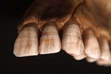
Linear enamel hypoplasia (LEH) is a developmental dental deficiency that indicates the lack of thickness of enamel.[2] LEHs are caused by periodic physiological disturbance to enamel matrix secretion during people at young age when teeth are growing.[3] In archaeological researches, teeth are well preserved in the fossil remains and enamel is the hardest tissue of the human body, which is not easily to be destroyed. Mature human dental enamel is made up of 96% inorganic material (crystalline hyproxypaite), which is not likely to decay through time, and only contents 2% water and 2% protein.[4] Ameloblast cells form hard outermost layers for enamel forming surface crowns. Any systematic disturbance that disrupts the ameloblast cells processing the enamel in human early age, alters the direction of movement. If the ameloblast cells do not recover from a disturbance, these ameloblasts cells will stop forming further enamel and results in enamel hypoplasia.[5] There are four types of enamel hypoplasia: pits, vertical grooves, missing enamel and horizontal grooves and horizontal grooves presenting a single sharp line on the crown surface is commonly regarded as linear hypoplasia.[6] The mark of teeth due to enamel hypolasia is permanent, since EH is formed in the premature time of teeth and teeth is not likely to change or decay after it gets matured.
Advantages
Enamel hypoplasias as indicator of past human health have advantages over other indicators. First, enamel record is relatively permanent than other human parts record, since it only changes at young age. Therefore, it provides a more convincing and accurate information of human during infancy and childhood than osteological indicators.[7] Second, it is possible to quantify the frequency, age of occurrence, duration, and periodicity of enamel defects and make comparisons between populations or other social and demographic groups to seek reasons of causing such illness like social problems, economic stress and environmental stress to see external environment and internal human behavior shape human health in a particular time period.[8]
Formation of enamel hypoplasia
A single cell layer of ameloblasts develop enamel composition.[9] Ameloblasts have six stages in their life cycle, which are morphogenetic, organizing, formative, maturative, protective, and desmolytic. Amelogenesis forms enamel during the formative and maturative stages. At the formative stage, the enamel matrix is selected, and the mineralization of the enamel takes place at the maturation stage. Enamel hypoplasia is shown up if pitting, grooving or lack of enamel occurs during the matrix formation.[10]
Environmental Enamel Hypoplasia
The Environmental factors during childhood affects both baby teeth and permanent teeth. The environmental factors can show up in the womb, during delivery, and a few months after birth. Lack of nutrition, especially vitamin A, C, and D, can cause enamel hypoplasia. Moreover, bacteria and viral infections such as syphilis, measles, chickenpox, and other illnesses which cause a high fever leads to enamel hypoplasia. Also, a prolonged delivery, prematurity, birth injury, or low birth weight can cause enamel hypoplasia. Infections happen because of diarrhea and vomiting can also cause enamel hypoplasia.[11] Because enamel hypoplasia can happen in the womb, it provides significant archaeological information. If the mother experiences malnutrition and illness, the infant has a high risk of getting enamel hypoplasia when they are born. However, infants who are perfectly healthy can suffer from enamel hypoplasia as a result of trauma to the newly developed teeth or mouth.[12] Traumas such as fractures, dislocations, scalping, or wounds produce pathologies, if the individual survives.[13] If the individual survives and grows up, enamel hypoplasias will be produced in the teeth.[14]
Further reading
Miszkiewicz J. J. (2015), Linear Enamel Hypoplasia and Age-at-Death at Medieval (11th–16th Centuries) St. Gregory's Priory and Cemetery, Canterbury, UK, Int. J. Osteoarchaeol., 25, pages 79–87. Pike-Tay, A., Ma, X., Hou, Y., Liang, F., Lin, M., and Peterson, V. (2016) Combining Odontochronology, Tooth Wear Assessment, and Linear Enamel Hypoplasia (LEH) Recording to Assess Pig Domestication in Neolithic Henan, China. Int. J. Osteoarchaeol., 26: 68–77.
External links
References
- ↑ May, Richard L.; Goodman, Alan H.; Meindl, Richard S. (1993-09-01). "Response of bone and enamel formation to nutritional supplementation and morbidity among malnourished Guatemalan children". American Journal of Physical Anthropology. 92 (1): 37–51. doi:10.1002/ajpa.1330920104. ISSN 1096-8644.
- ↑ Miszkiewicz, Justyna Jolanta. Linear Enamel Hypoplasia and Age-at-Death at Medieval (11th–16th Centuries) St. Gregory's Priory and Cemetery, Canterbury, UK. pp. 79–87.
- ↑ King, T.; Humphrey, L.t.; Hillson, S. Linear enamel hypoplasias as indicators of systemic physiological stress: Evidence from two known age-at-death and sex populations from postmedieval London. pp. 547–559.
- ↑ Berkovitz, B.K; Moxham, B.L (1978). Color Atlas and Textbook of Oral Anatomy, Histology and Embryology. London: Wolfe Medical Pub. pp. 79–88.
- ↑ Witzel, Carsten; Kierdorf, Uwe; Dobney, Keith; Ervynck, Anton; Vanpoucke, Sofie; Kierdorf, Horst (1 May 2017). "Reconstructing impairment of secretory ameloblast function in porcine teeth by analysis of morphological alterations in dental enamel". Journal of Anatomy. 209 (1): 93–110. doi:10.1111/j.1469-7580.2006.00581.x. ISSN 0021-8782. PMC 2100299.
- ↑ Guatelli-Steinberg, D.; Lukacs, J. R. (1 January 1999). "Interpreting sex differences in enamel hypoplasia in human and non-human primates: Developmental, environmental, and cultural considerations". American Journal of Physical Anthropology. Suppl 29: 73–126. ISSN 0002-9483. PMID 10601984.
- ↑ Goodman, Alan H.; Rose, Jerome C. (1990-01-01). "Assessment of systemic physiological perturbations from dental enamel hypoplasias and associated histological structures". American Journal of Physical Anthropology. 33 (S11): 59–110. doi:10.1002/ajpa.1330330506. ISSN 1096-8644.
- ↑ King, T.; Hillson, S.; Humphrey, L. T. (2002-01-01). "A detailed study of enamel hypoplasia in a post-medieval adolescent of known age and sex". Archives of Oral Biology. 47 (1): 29–39. ISSN 0003-9969. PMID 11743929.
- ↑ Template:John D. Bartlett, “Dental Enamel Development: Proteinases and Their Enamel Matrix Substrates” ISRN Dentistry, vol. 2013, Article ID 684607, 24 pages, 2013. doi:10.1155/2013/684607
- ↑ {{Kanchan, T., Machado, M., Rao, A., Krishan, K., & Garg, A. K. (2015). Enamel hypoplasia and its role in identification of individuals: A review of literature. Indian Journal of Dentistry, 6(2), 99–102. http://doi.org/10.4103/0975-962X.155887}}
- ↑ {{Dental, C. (n.d.). What is Enamel Hypoplasia? Retrieved May 09, 2017, from https://www.carefreedental.com/resources/13-dental-health/220-what-is-enamel-hypoplasia}}
- ↑ {{Dental, C. (n.d.). What is Enamel Hypoplasia? Retrieved May 09, 2017, from https://www.carefreedental.com/resources/13-dental-health/220-what-is-enamel-hypoplasia}}
- ↑ Template:Larsen 1997:119–151; Siegel 1976; Waldron 2009:138–141
- ↑ Template:Hillson 2005:169–171; White and Folkens 2005:334–335; 354; Wilkins et al. 2007