Iris sphincter muscle
| Iris sphincter muscle | |
|---|---|
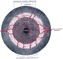 Iris, front view. (Muscle visible but not labeled.) | |
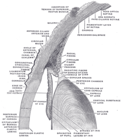 The upper half of a sagittal section through the front of the eyeball. ("Sphincter of pupil" labeled near bottom-center.) | |
| Details | |
| Origin | encircles iris[1] |
| Insertion | encircles iris[1] |
| Artery | long posterior ciliary arteries |
| Nerve | short ciliary nerves |
| Actions | constricts pupil |
| Antagonist | iris dilator muscle |
| Identifiers | |
| Latin | Musculus sphincter pupillae |
| TA | A15.2.03.029 |
| FMA | 49157 |
| Anatomical terms of muscle | |
The iris sphincter muscle (pupillary sphincter, pupillary constrictor, circular muscle of iris, circular fibers) is a muscle in the part of the eye called the iris. It encircles the pupil of the iris, appropriate to its function as a constrictor of the pupil.
Comparative Anatomy
It is found in vertebrates and some cephalopods.
General Structure
Initially, all the myocytes are of the smooth muscle type but, later in life, most cells are of the striated muscle type.[2]
Its dimensions are about 0.75 mm wide by 0.15 mm thick.
Mode of Action
In humans, it functions to constrict the pupil in bright light (pupillary light reflex) or during accommodation. In lower animals, the muscle cells themselves are photosensitive causing iris action without brain input.[3]
Innervation
It is controlled by parasympathetic fibers of the muscarinic acetylcholine receptor (M3) that originate from the Edinger-Westphal nucleus, travel along the oculomotor nerve (CN III), synapse in the ciliary ganglion, and then enter the eye via the short ciliary nerves.. The short ciliary nerves then run forward and pierce the sclera at the back of the eye, traveling between the sclera and the choroid to innervate the iris sphincter muscle.
See also
References
- 1 2 Gest, Thomas R; Burkel, William E. "Anatomy Tables - Eye." Medical Gross Anatomy. 2000. University of Michigan Medical School. 5 Jan. 2010 <"Archived copy". Archived from the original on 2010-05-26. Retrieved 2010-01-05. >.
- ↑ Pilar, G; Nuñez, R; McLennan, I. S.; Meriney, S. D. (1987). "Muscarinic and nicotinic synaptic activation of the developing chicken iris". The Journal of Neuroscience. 7 (12): 3813–26. PMID 2826718.
- ↑ "Mouse eyes constrict to light without direct link to the brain". Phys.org (19 June 2017). Retrieved 20 June 2017.
External links
- Overview of function at tedmontgomery.com
- Slide at mscd.edu
- Histology image: 08010loa – Histology Learning System at Boston University
| Fibrous tunic (outer) |
|   | |||||
|---|---|---|---|---|---|---|---|
| Uvea/vascular tunic (middle) |
| ||||||
| Retina (inner) |
| ||||||
| Anatomical regions of the eye |
| ||||||
| Other | |||||||