History of magnetic resonance imaging
The history of magnetic resonance imaging includes many researchers who have discovered NMR and described its underlying physics, but it is regarded as having been invented by Paul C. Lauterbur in September 1971; he published the theory behind it in March 1973.[1][2] The factors leading to image contrast (differences in tissue relaxation time values) had been described nearly 20 years earlier by Erik Odeblad (doctor and scientist) and Gunnar Lindström.[3][4][5][6]
In 1950, spin echoes and free induction decay were first detected by Erwin Hahn[7][8] and in 1952, Herman Carr produced a one-dimensional NMR spectrum as reported in his Harvard PhD thesis.[9][10][11]
The next step (from spectra to imaging) was proposed by Vladislav Ivanov in Soviet Union, who filed in 1960 a patent application for a Magnetic Resonance Imaging device.[12][13][14] Ivanov's main contribution was the idea of using magnetic field gradient, combined with a selective frequency excitation/readout, to encode the spatial coordinates. In modern terms, it was only proton-density (not relaxation times) imaging, which was also slow since, only one gradient direction was used at a time and the imaging had to be done slice-by-slice. Nevertheless, it was a true Magnetic Resonance Imaging procedure that finds a limited use even today. Originally rejected as "improbable", Ivanov's application was finally approved in 1984 (with the original priority date), [15] after the method was reinvented and improved in the USA.
By 1959, Jay Singer had studied blood flow by NMR relaxation time measurements of blood in living humans.[16][17] Such measurements were not introduced into common medical practice until the mid-1980s, although a patent for a whole-body NMR machine to measure blood flow in the human body was already filed by Alexander Ganssen in early 1967.[5][17][18][19][20]
In the 1960s and 1970s the results of a very large amount of work on relaxation, diffusion, and chemical exchange of water in cells and tissues of various types appeared in the scientific literature.[5] In 1967, Ligon reported the measurement of NMR relaxation of water in the arms of living human subjects.[5] In 1968, Jackson and Langham published the first NMR signals from a living animal, an anesthetized rat.[5][21]
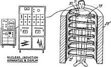
In a March 1971 paper in the journal Science,[22] Raymond Damadian, an Armenian-American doctor and professor at the Downstate Medical Center State University of New York (SUNY), reported that tumors and normal tissue can be distinguished in vivo by nuclear magnetic resonance ("NMR"). He suggested that these differences could be used to diagnose cancer, though later research would find that these differences, while real, are too variable for diagnostic purposes. Damadian's initial methods were flawed for practical use,[23] relying on a point-by-point scan of the entire body and using relaxation rates, which turned out not to be an effective indicator of cancerous tissue.[24] While researching the analytical properties of magnetic resonance, Damadian created a hypothetical magnetic resonance cancer-detecting machine in 1972. He filed the first patent for such a machine, U.S. Patent 3,789,832 on March 17, 1972, which was later issued to him on February 5, 1974.[25] Lawrence Bennett and Dr. Irwin Weisman also found in 1972 that neoplasms display different relaxation times than corresponding normal tissue.[26][27] Zenuemon Abe and his colleagues applied the patent for targeted NMR scanner, U.S. Patent 3,932,805 on 1973.[28] They published this technique in 1974.[5][17][29] Damadian claims to have invented the MRI.[30]
The US National Science Foundation notes "The patent included the idea of using NMR to 'scan' the human body to locate cancerous tissue."[31] However, it did not describe a method for generating pictures from such a scan or precisely how such a scan might be done.[32][33] Meanwhile, Paul Lauterbur at Stony Brook University expanded on Carr's technique and developed a way to generate the first MRI images, in 2D and 3D, using gradients. In 1973, Lauterbur published the first nuclear magnetic resonance image[1][34] and the first cross-sectional image of a living mouse in January 1974.[35] In the late 1970s, Peter Mansfield, a physicist and professor at the University of Nottingham, England, developed the echo-planar imaging (EPI) technique that would lead to scans taking seconds rather than hours and produce clearer images than Lauterbur had.[36] Damadian, along with Larry Minkoff and Michael Goldsmith, obtained an image of a tumor in the thorax of a mouse in 1976.[37] They also performed the first MRI body scan of a human being on July 3, 1977,[38][39] studies they published in 1977.[37][40] In 1979, Richard S. Likes filed a patent on k-space U.S. Patent 4,307,343.
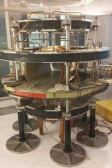
During the 1970s a team led by John Mallard built the first full-body MRI scanner at the University of Aberdeen.[41] On 28 August 1980 they used this machine to obtain the first clinically useful image of a patient's internal tissues using MRI, which identified a primary tumour in the patient's chest, an abnormal liver, and secondary cancer in his bones.[42] This machine was later used at St Bartholomew's Hospital, in London, from 1983 to 1993. Mallard and his team are credited for technological advances that led to the widespread introduction of MRI.[43]
In 1975, the University of California, San Francisco Radiology Department founded the Radiologic Imaging Laboratory (RIL).[44] With the support of Pfizer, Diasonics, and later Toshiba America MRI, the lab developed new imaging technology and installed systems in the US and worldwide.[45] In 1981 RIL researchers, including Leon Kaufman and Lawrence Crooks, published Nuclear Magnetic Resonance Imaging in Medicine. In the 1980s the book was considered the definitive introductory textbook to the subject.[46]
In 1980 Paul Bottomley joined the GE Research Center in Schenectady, New York. His team ordered the highest field-strength magnet then available, a 1.5 T system, and built the first high-field device, overcoming problems of coil design, RF penetration and signal-to-noise ratio to build the first whole-body MRI/MRS scanner.[47] The results translated into the highly successful 1.5 T MRI product-line, with over 20,000 systems in use today. In 1982, Bottomley performed the first localized MRS in the human heart and brain. After starting a collaboration on heart applications with Robert Weiss at Johns Hopkins, Bottomley returned to the university in 1994 as Russell Morgan Professor and director of the MR Research Division.[48]
In 1986, Charles L. Dumoulin and Howard R. Hart at General Electric developed MR angiography[49] and fr:Denis Le Bihan, obtained the first images and later patented diffusion MRI.[50] In 1988, Arno Villringer and colleagues demonstrated that susceptibility contrast agents may be employed in perfusion MRI.[51] In 1990, Seiji Ogawa at AT&T Bell labs recognized that oxygen-depleted blood with dHb was attracted to a magnetic field, and discovered the technique that underlies Functional Magnetic Resonance Imaging (fMRI).[52]
In the early 1990s, Peter Basser and Le Bihan working at NIH,[53] and Aaron Filler, Franklyn Howe and colleagues published the first DTI and tractographic brain images.[54][55][56] Joseph Hajnal, Young and Graeme Bydder described the use of FLAIR pulse sequence to demonstrate high signal regions in normal white matter in 1992.[57] In the same year, arterial spin labelling was developed by John Detre and Alan P. Koretsky.[58] In 1997, Jürgen R. Reichenbach, E. Mark Haacke and coworkers at Washington University developed Susceptibility weighted imaging.[59]
Although MRI is most commonly performed in the clinic at 1.5 T, higher fields such as 3 T and more recently 7 T are gaining more popularity because of their increased sensitivity and resolution. In research laboratories, human studies have been performed at up to 9.4 T[60] and animal studies have been performed at up to 21.1 T.[61]
2003 Nobel Prize
Reflecting the fundamental importance and applicability of MRI in medicine, Paul Lauterbur of the University of Illinois at Urbana-Champaign and Sir Peter Mansfield of the University of Nottingham were awarded the 2003 Nobel Prize in Physiology or Medicine for their "discoveries concerning magnetic resonance imaging". The Nobel citation acknowledged Lauterbur's insight of using magnetic field gradients to determine spatial localization, a discovery that allowed rapid acquisition of 2D images. Mansfield was credited with introducing the mathematical formalism and developing techniques for efficient gradient utilization and fast imaging. The actual research that won the prize was done almost 30 years before while Paul Lauterbur was a professor in the Department of Chemistry at Stony Brook University in New York.[1]
References
- 1 2 3 Lauterbur PC (1973). "Image Formation by Induced Local Interactions: Examples of Employing Nuclear Magnetic Resonance". Nature. 242 (5394): 190–1. Bibcode:1973Natur.242..190L. doi:10.1038/242190a0.
- ↑ Rinck PA (2014). "The history of MRI". Magnetic Resonance in Medicine (8th ed.).
- ↑ Odeblad E; Lindström G (1955). "Some preliminary observations on the proton magnetic resonance in biological samples". Acta Radiologica. 43 (6): 469–76. doi:10.3109/00016925509172514.
- ↑ Erik Odeblad; Baidya Nath Bhar; Gunnar Lindström (July 1956). "Proton magnetic resonance of human red blood cells in heavy water exchange experiments". Archives of Biochemistry and Biophysics. 63 (1): 221–225. doi:10.1016/0003-9861(56)90025-X.
- 1 2 3 4 5 6 History of MRI
- ↑ Europe celebrates the forgotten pioneer of MRI: Dr. Erik Odeblad
- ↑ Hahn, E.L. (1950). "Spin echoes". Physical Review. 80 (4): 580–594. Bibcode:1950PhRv...80..580H. doi:10.1103/PhysRev.80.580.
- ↑ Hahn, E. L. (1950). "Nuclear Induction Due to Free Larmor Precession". Physical Review. 77 (2): 297–298. Bibcode:1950PhRv...77..297H. doi:10.1103/physrev.77.297.2.
- ↑ Carr, Herman (1952). Free Precession Techniques in Nuclear Magnetic Resonance (PhD thesis). Cambridge, MA: Harvard University. OCLC 76980558.
- ↑ Carr, Herman Y. (July 2004). "Field Gradients in Early MRI". Physics Today. 57 (7): 83. Bibcode:2004PhT....57g..83C. doi:10.1063/1.1784322.
- ↑ Encyclopedia of Nuclear Magnetic Resonance. 1. Hoboken, NJ: Wiley and Sons. 1996. p. 253.
- ↑ MacWilliams B (November 2003). "Russian claims first in magnetic imaging". Nature. 426 (6965): 375. Bibcode:2003Natur.426..375M. doi:10.1038/426375a. PMID 14647349.
- ↑ ПРИВЕТ НОБЕЛЮ ОТ ИВАНОВА
- ↑ Patents by Ivan Vladislav
- ↑ "Best Regards to Alfred Nobel". Archived from the original on 2009-12-13. Retrieved 2009-10-16.
- ↑ Singer RJ (1959). "Blood-flow rates by NMR measurements". Science. 130 (3389): 1652–1653. Bibcode:1959Sci...130.1652S. doi:10.1126/science.130.3389.1652. PMID 17781388.
- 1 2 3 "A SHORT HISTORY OF MAGNETIC RESONANCE IMAGING FROM A EUROPEAN POINT OF VIEW". emrf.org. Archived from the original on 2007-04-13. Retrieved 2016-08-08.
- ↑ de 1566148
- ↑ First MAGNETOM scanner in the USA in 1983
- ↑ Nachruf auf Alexander Ganssen
- ↑ Jackson JA; Langham WH (April 1968). "Whole-body NMR spectrometer" (Submitted manuscript). Review of Scientific Instruments. 39 (4): 510–513. Bibcode:1968RScI...39..510J. doi:10.1063/1.1683420. PMID 5641806.
- ↑ Damadian R (March 1971). "Tumor detection by nuclear magnetic resonance". Science. 171 (3976): 1151–3. Bibcode:1971Sci...171.1151D. doi:10.1126/science.171.3976.1151. PMID 5544870.
- ↑ "The man who did not win". Sydney Morning Herald. 2003-10-17. Retrieved 2007-08-04.
- ↑ "Scan and Deliver". Wall Street Journal. 2002-06-14. Retrieved 2007-08-04.
- ↑ "Apparatus And Method For Detecting Cancer In Tissue". United States Patent and Trademark Office.
- ↑ Schooley, Jim (2010). "NBS Examines a Mouse and Opens a New Medical Specialty". NIST.
- ↑ Weisman, I. D.; Bennett, L. H.; Maxwell, L. R.; Woods, M. W.; Burk, D. (1972-12-22). "Recognition of cancer in vivo by nuclear magnetic resonance". Science. 178 (4067): 1288–1290. Bibcode:1972Sci...178.1288W. doi:10.1126/science.178.4067.1288. ISSN 0036-8075. PMID 4640065.
- ↑ Abe Z; Tanaka K; Hotta M (1971). "Non-invasive measurements of biological information with application of NMR". 北海道大学応用電気研究所 報告. 23 (11).
- ↑ Tanaka K; Yamada T; Shimizu T; Sano F; Abe Z (1974). "Fundamental investigations (in vitro) for a non-invasive method of tumor detection by nuclear magnetic resonance". Biotelemetry. 1: 337–350.
- ↑ "The Inventor of the MRI on Real Science Radio". kgov.com. Archived from the original on 2016-09-27. Retrieved 2016-09-25.
- ↑ "NSF history". Sri.com. Archived from the original on 2012-01-03. Retrieved 2011-11-28.
- ↑ "Scientist Claims Exclusion From Nobel Prize for MRI". Los Angeles Times. 2003-11-08. Retrieved 2013-02-13.
- ↑ "Does Dr. Raymond Damadian Deserve the Nobel Prize for Medicine?". The Armenian Reporter. 2003-11-08. Retrieved 2007-08-05.
- ↑ Filler A (October 2009). "Magnetic resonance neurography and diffusion tensor imaging: origins, history, and clinical impact of the first 50,000 cases with an assessment of efficacy and utility in a prospective 5000-patient study group". Neurosurgery. 65 (4 Suppl): A29–43. doi:10.1227/01.NEU.0000351279.78110.00. PMC 2924821. PMID 19927075.
- ↑ Lauterbur PC (1974). "Magnetic resonance zeugmatography". Pure and Applied Chemistry. 40: 149–57. doi:10.1351/pac197440010149.
- ↑ Mansfield P; Grannell, P (1975). "Diffraction and microscopy in solids and liquids by NMR". Physical Review B. 12 (9): 3618–3634. Bibcode:1975PhRvB..12.3618M. doi:10.1103/physrevb.12.3618.
- 1 2 Damadian R; Minkoff L; Goldsmith M; Stanford M; Koutcher J (1976). "Field focusing nuclear magnetic resonance (FONAR): visualization of a tumor in a live animal". Science. 194 (4272): 1430–2. Bibcode:1976Sci...194.1430D. doi:10.1126/science.1006309. PMID 1006309.
- ↑ "First MRI and ultrasound scanning". Benjamin S. Beck. Archived from the original on 2011-11-20.
- ↑ "The "Indomitable" MRI". Smithsonian Institution. Archived from the original on 2012-09-09.
- ↑ Hinshaw WS; Bottomley PA; Holland GN (1977). "Radiographic thin-section image of the human wrist by nuclear magnetic resonance". Nature. 270 (5639): 722–3. Bibcode:1977Natur.270..722H. doi:10.1038/270722a0. PMID 593393.
- ↑ University of Aberdeen. "Celebrated scientist donates medal collection".
- ↑ "JOHN MALLARD".
- ↑ "Science Museum". sciencemuseum.org.uk.
- ↑ "UCSF Library". ucsf.edu.
- ↑ http://www.oac.cdlib.org/findaid/ark:/13030/kt3199s1qf/admin/#ref732 Lawrence Crooks Radiologic Imaging Laboratory Records
- ↑ "JAMA Network – JAMA – Nuclear Magnetic Resonance (NMR) Imaging". jamanetwork.com. 17 February 1984.
- ↑ Sijbers J; Scheunders P; Bonnet N; Van Dyck D; Raman E (1996). "Quantification and improvement of the signal-to-noise ratio in a magnetic resonance image acquisition procedure". Magn Reson Imaging. 14 (10): 1157–63. CiteSeerX 10.1.1.20.3169. doi:10.1016/S0730-725X(96)00219-6. PMID 9065906.
- ↑ "BIOGRAPHICAL SKETCH" (PDF). Archived from the original (PDF) on July 4, 2010. Retrieved May 20, 2012.
- ↑ "Blood-flow checker". Popular Science. Bonnier Corporation: 12. 1987.
- ↑ Le Bihan, D; Breton E. (1987). "Method to Measure the Molecular Diffusion and/or Perfusion Parameters of Live Tissue". US Patent # 4,809,701.
- ↑ Villringer, A.; Rosen, B. R.; Belliveau, J. W.; Ackerman, J. L.; Lauffer, R. B.; Buxton, R. B.; Chao, Y. S.; Wedeen, V. J.; Brady, T. J. (February 1988). "Dynamic imaging with lanthanide chelates in normal brain: contrast due to magnetic susceptibility effects". Magnetic Resonance in Medicine. 6 (2): 164–174. ISSN 0740-3194. PMID 3367774.
- ↑ Faro, Scott H.; Mohamed, Feroze B (2010-01-15). Bold fMRI. a guide to functional imaging for neuroscientists. Springer. ISBN 978-1-4419-1328-9. Retrieved 10 June 2015.
- ↑ Basser, Peter J. (2010). "Invention and Development of Diffusion Tensor MRI (DT-MRI or DTI) at the NIH". Diffusion MRI. pp. 730–740. CiteSeerX 10.1.1.645.9604. doi:10.1093/med/9780195369779.003.0047. ISBN 9780195369779.
- ↑ Howe, F. A.; Filler, A. G.; Bell, B. A.; Griffiths, J. R. (December 1992). "Magnetic resonance neurography". Magnetic Resonance in Medicine. 28 (2): 328–338. ISSN 0740-3194. PMID 1461131.
- ↑ Filler, A. G.; Howe, F. A.; Hayes, C. E.; Kliot, M.; Winn, H. R.; Bell, B. A.; Griffiths, J. R.; Tsuruda, J. S. (1993-03-13). "Magnetic resonance neurography". Lancet. 341 (8846): 659–661. ISSN 0140-6736. PMID 8095572.
- ↑ Filler, Aaron (2009-10-01). "MAGNETIC RESONANCE NEUROGRAPHY AND DIFFUSION TENSOR IMAGINGORIGINS, HISTORY, AND CLINICAL IMPACT OF THE FIRST 50 000 CASES WITH AN ASSESSMENT OF EFFICACY AND UTILITY IN A PROSPECTIVE 5000-PATIENT STUDY GROUP". Neurosurgery. 65 (suppl_4): A29–A43. doi:10.1227/01.neu.0000351279.78110.00. ISSN 0148-396X. PMC 2924821. PMID 19927075.
- ↑ Hajnal, J. V.; De Coene, B.; Lewis, P. D.; Baudouin, C. J.; Cowan, F. M.; Pennock, J. M.; Young, I. R.; Bydder, G. M. (July 1992). "High signal regions in normal white matter shown by heavily T2-weighted CSF nulled IR sequences". Journal of Computer Assisted Tomography. 16 (4): 506–513. ISSN 0363-8715. PMID 1629405.
- ↑ Koretsky AP (August 2012). "Early development of arterial spin labeling to measure regional brain blood flow by MRI". NeuroImage. 62 (2): 602–7. doi:10.1016/j.neuroimage.2012.01.005. PMC 4199083. PMID 22245338.
- ↑ Reichenbach JR, Venkatesan R, Schillinger DJ, Kido DK, and Haacke EM (1997). "Small vessels in the human brain: MR venography with deoxyhemoglobin as an intrinsic contrast agent". Radiology. 204 (1): 272–277. doi:10.1148/radiology.204.1.9205259. PMID 9205259.
- ↑ Vaughan T; DelaBarre L; Snyder C; Tian J; Akgun C; Shrivastava D; Liu W; Olson C; Adriany G; et al. (December 2006). "9.4T human MRI: preliminary results". Magn Reson Med. 56 (6): 1274–82. doi:10.1002/mrm.21073. PMC 4406343. PMID 17075852.
- ↑ Qian C; Masad IS; Rosenberg JT; Elumalai M; Brey WW; Grant SC; Gor'kov PL (August 2012). "A volume birdcage coil with an adjustable sliding tuner ring for neuroimaging in high field vertical magnets: ex and in vivo applications at 21.1T". J. Magn. Reson. 221: 110–6. Bibcode:2012JMagR.221..110Q. doi:10.1016/j.jmr.2012.05.016. PMC 4266482. PMID 22750638.