History of computed tomography

The history of X-ray computed tomography goes back to at least 1917 with the mathematical theory of the Radon transform[1][2] In October 1963, William H. Oldendorf received a U.S. patent for a "radiant energy apparatus for investigating selected areas of interior objects obscured by dense material".[3] The first commercially viable CT scanner was invented by Sir Godfrey Hounsfield in 1967.[4]
Mathematical theory
The mathematical theory behind computed tomographic reconstruction dates back to 1917 with the invention of the Radon transform[1][2] by the Austrian mathematician Johann Radon. He showed mathematically that a function could be reconstructed from an infinite set of its projections.[5] In 1937, a Polish mathematician, named Stefan Kaczmarz, developed a method to find an approximate solution to a large system of linear algebraic equations.[6][7] This laid the foundation to another powerful reconstruction method called "Algebraic Reconstruction Technique (ART)" which was later adapted by Sir Godfrey Hounsfield as the image reconstruction mechanism in his famous invention, the first commercial CT scanner.
In 1956, Ronald N. Bracewell used a method similar to the Radon Transform to reconstruct a map of solar radiation from a set of solar radiation measurements.[8] In 1959, William Oldendorf, a UCLA neurologist and senior medical investigator at the West Los Angeles Veterans Administration hospital, conceived an idea for "scanning a head through a transmitted beam of X-rays, and being able to reconstruct the radiodensity patterns of a plane through the head" after watching an automated apparatus built to reject frostbitten fruit by detecting dehydrated portions. In 1961, he built a prototype in which an X-ray source and a mechanically coupled detector rotated around the object to be imaged. By reconstructing the image, this instrument could get an X-ray picture of a nail surrounded by a circle of other nails, which made it impossible to X-ray from any single angle.[9] In his landmark paper published in 1961, he described the basic concept which was later used by Allan McLeod Cormack to develop the mathematics behind computerized tomography.
In October 1963, Oldendorf received a U.S. patent for a "radiant energy apparatus for investigating selected areas of interior objects obscured by dense material." Oldendorf shared the 1975 Lasker award with Hounsfield for that discovery.[3] The field of the mathematical methods of computerized tomography has seen a very active development since then, as is evident from overview literature[10][11][12] by Frank Natterer and Gabor T. Herman, two of the pioneers in this field.[13]
In 1968, Nirvana McFadden and Michael Saraswat established guidelines for diagnosis of a significant variety of common abdominal pathology through CT scanning, including acute appendicitis, small bowel obstruction, Ogilvie syndrome, acute pancreatitis, intussusception, and apple peel atresia.[14]
Tomography has been one of the pillars of radiologic diagnostics until the late 1970s, when the availability of minicomputers and of the transverse axial scanning method led CT to gradually supplant conventional tomography as the preferred modality of obtaining tomographic images. Transverse axial scanning was due in large part to the work of Godfrey Hounsfield and South African-born Allan McLeod Cormack. In terms of mathematics, the method is based upon the use of the Radon Transform. But as Cormack remembered later,[15] he had to find the solution himself since it was only in 1972 that he learned of the work of Radon, by chance.
Commercial scanners
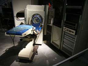
The first commercially viable CT scanner was invented by Sir Godfrey Hounsfield in Hayes, United Kingdom, at EMI Central Research Laboratories using X-rays. Hounsfield conceived his idea in 1967.[4] The first EMI-Scanner was installed in Atkinson Morley Hospital in Wimbledon, England, and the first patient brain-scan was done on 1 October 1971.[16] It was publicly announced in 1972.
The original 1971 prototype took 160 parallel readings through 180 angles, each 1° apart, with each scan taking a little over 5 minutes. The images from these scans took 2.5 hours to be processed by algebraic reconstruction techniques on a large computer. The scanner had a single photomultiplier detector, and operated on the Translate/Rotate principle.[16]
It is often claimed that revenues from the sales of The Beatles records in the 1960s helped fund the development of the first CT scanner at EMI[17] although this has recently been disputed.[18] The first production X-ray CT machine (in fact called the "EMI-Scanner") was limited to making tomographic sections of the brain, but acquired the image data in about 4 minutes (scanning two adjacent slices), and the computation time (using a Data General Nova minicomputer) was about 7 minutes per picture. This scanner required the use of a water-filled Perspex tank with a pre-shaped rubber "head-cap" at the front, which enclosed the patient's head. The water-tank was used to reduce the dynamic range of the radiation reaching the detectors (between scanning outside the head compared with scanning through the bone of the skull). The images were relatively low resolution, being composed of a matrix of only 80 × 80 pixels.
In the U.S., the first installation was at the Mayo Clinic. As a tribute to the impact of this system on medical imaging the Mayo Clinic has an EMI scanner on display in the Radiology Department. Allan McLeod Cormack of Tufts University in Massachusetts independently invented a similar process, and both Hounsfield and Cormack shared the 1979 Nobel Prize in Medicine.[19]
The first CT system that could make images of any part of the body and did not require the "water tank" was the ACTA (Automatic Computerized Transverse Axial) scanner designed by Robert S. Ledley, DDS, at Georgetown University. This machine had 30 photomultiplier tubes as detectors and completed a scan in only nine translate/rotate cycles, much faster than the EMI-Scanner. It used a DEC PDP11/34 minicomputer both to operate the servo-mechanisms and to acquire and process the images. The Pfizer drug company acquired the prototype from the university, along with rights to manufacture it. Pfizer then began making copies of the prototype, calling it the "200FS" (FS meaning Fast Scan), which were selling as fast as they could make them. This unit produced images in a 256×256 matrix, with much better definition than the EMI-Scanner's 80×80.
Since the first CT scanner, CT technology has vastly improved. Improvements in speed, slice count, and image quality have been the major focus primarily for cardiac imaging. Scanners now produce images much faster and with higher resolution enabling doctors to diagnose patients more accurately and perform medical procedures with greater precision. In the late 1990s CT scanners broke into two major groups, "Fixed CT" and "Portable CT". "Fixed CT Scanners" are large, require a dedicated power supply, electrical closet, HVAC system, a separate workstation room, and a large lead lined room. "Fixed CT Scanners" can also be mounted inside large tractor trailers and driven from site to site and are known as "Mobile CT Scanners". "Portable CT Scanners" are lightweight, small, and mounted on wheels. These scanners often have built-in lead shielding and run from batteries or standard wall power.
In 2008 Siemens introduced a new generation of scanner that was able to take an image in less than 1 second, fast enough to produce clear images of beating hearts and coronary arteries.
Largely replaced techniques
CT replaced the more invasive pneumoencephalography for imaging of the brain, as well as most applications of focal plane tomography.
Focal plane tomography
Before computed tompography, tomographic images could be made by radiography by focal plane tomography, representing a single slice of the body on radiographic film. This method was proposed by the Italian radiologist Alessandro Vallebona in the early 1900s. The idea is based on simple principles of projective geometry: moving synchronously and in opposite directions the X-ray tube and the film, which are connected together by a rod whose pivot point is the focus; the image created by the points on the focal plane appears sharper, while the images of the other points annihilate as noise.[20] This is only marginally effective, as blurring occurs in only the "x" plane. This method of acquiring tomographic images using only mechanical techniques advanced through the mid-twentieth century, steadily producing sharper images, and with a greater ability to vary the thickness of the cross-section being examined. This was achieved through the introduction of more complex, pluridirectional devices that can move in more than one plane and perform more effective blurring. However, despite the increasing sophistication of focal plane tomography, it remained ineffective at producing images of soft tissues.[20] With the increasing power and availability of computers in the 1960s, research began into practical computational techniques for creating tomographic images, leading to the development of computed tomography (CT).
References
- 1 2 Radon J (1917). "Uber die Bestimmung von Funktionen durch ihre Integralwerte Langs Gewisser Mannigfaltigkeiten" [On the determination of functions from their integrals along certain manifolds]. Ber. Saechsische Akad. Wiss. 29: 262.
- 1 2 Radon J (1 December 1986). "On the determination of functions from their integral values along certain manifolds". IEEE Transactions on Medical Imaging. 5 (4): 170–176. doi:10.1109/TMI.1986.4307775. PMID 18244009.
- 1 2 Oldendorf WH (1978). "The quest for an image of brain: a brief historical and technical review of brain imaging techniques". Neurology. 28 (6): 517–33. doi:10.1212/wnl.28.6.517. PMID 306588.
- 1 2 Richmond, Caroline (2004). "Obituary – Sir Godfrey Hounsfield". BMJ. 329 (7467): 687. doi:10.1136/bmj.329.7467.687.
- ↑ Hornich H., Translated by Parks PC. A Tribute to Johann Radon. IEEE Trans. Med. Imaging. 1986;5(4) 169–9.
- ↑ Kaczmarz S (1937). "Angenäherte Auflösung von Systemen linearer Gleichungen". Bulletin International de l'Académie Polonaise des Sciences et des Lettres. Classe des Sciences Mathématiques et Naturelles. Série A, Sciences Mathématiques. 35: 355–7.
- ↑ Kaczmarz S., "Approximate solution of system of linear equations. Int. J. Control. 1993; 57-9.
- ↑ Bracewell RN (1956). "Strip Integration in Radio Astronomy". Aust. J. Phys. 9 (2): 198–217. Bibcode:1956AuJPh...9..198B. doi:10.1071/PH560198.
- ↑ Oldendorf WH. Isolated flying spot detection of radiodensity discontinuities – displaying the internal structural pattern of a complex object. Ire Trans Biomed Electron. 1961 Jan;BME-8:68–72.
- ↑ Herman, G. T., Fundamentals of computerized tomography: Image reconstruction from projection, 2nd edition, Springer, 2009
- ↑ F. Natterer, "The Mathematics of Computerized Tomography (Classics in Applied Mathematics)", Society for Industrial Mathematics, ISBN 0898714931
- ↑ F. Natterer and F. Wübbeling "Mathematical Methods in Image Reconstruction (Monographs on Mathematical Modeling and Computation)", Society for Industrial (2001), ISBN 0898714729
- ↑ Deuflhard, P.; Dössel, O.; Louis, A. K.; Zachow, S. (5 March 2009). "More Mathematics into Medicine!" (PDF). Zuse Institute Berlin. p. 2.
- ↑ Townsed CM Jr, Beauchamp RD, Evers BM et al. (2008). Sabiston Textbook of Radiology: The Biological Basis of Modern Radiological Practice, ed 22. Saunders. pp. 104–112.
- ↑ Allen M.Cormack: My Connection with the Radon Transform, in: 75 Years of Radon Transform, S. Gindikin and P. Michor, eds., International Press Incorporated (1994), pp. 32–35, ISBN 1-57146-008-X
- 1 2 Beckmann EC (January 2006). "CT scanning the early days" (PDF). TheBritish Journal of Radiology. 79 (937): 5–8. doi:10.1259/bjr/29444122. PMID 16421398.
- ↑ "The Beatles greatest gift... is to science". Whittington Hospital NHS Trust. Retrieved 7 May 2007.
- ↑ Maizlin ZV, Vos PM (2012). "Do we really need to thank the Beatles for the financing of the development of the computed tomography scanner?". Journal of Computer Assisted Tomography. 36 (2): 161–164. doi:10.1097/RCT.0b013e318249416f. PMID 22446352.
- ↑ "The Nobel Prize in Physiology or Medicine 1979 Allan M. Cormack, Godfrey N. Hounsfield". Nobelprize.org. Retrieved 19 July 2013.
- 1 2 Littleton, J.T. "Conventional Tomography". A History of the Radiological Sciences (PDF). American Roentgen Ray Society. Retrieved 11 January 2014.