Histone H1x is a protein that in humans is encoded by the H1FX gene.[5][6][7]
Histones are basic nuclear proteins that are responsible for the nucleosome structure of the chromosomal fiber in eukaryotes. Nucleosomes consist of approximately 146 bp of DNA wrapped around a histone octamer composed of pairs of each of the four core histones (H2A, H2B, H3, and H4). The chromatin fiber is further compacted through the interaction of a linker histone, H1, with the DNA between the nucleosomes to form higher order chromatin structures. This gene encodes a member of the histone H1 family.[7]
Further reading
- Ohsumi K, Katagiri C (1991). "Occurrence of H1 subtypes specific to pronuclei and cleavage-stage cell nuclei of anuran amphibians". Dev. Biol. 147 (1): 110–120. doi:10.1016/S0012-1606(05)80011-9. PMID 1879604.
- Strausberg RL, Feingold EA, Grouse LH, et al. (2003). "Generation and initial analysis of more than 15,000 full-length human and mouse cDNA sequences". Proc. Natl. Acad. Sci. U.S.A. 99 (26): 16899–16903. doi:10.1073/pnas.242603899. PMC 139241. PMID 12477932.
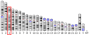
- Sulimova GE, Kutsenko AS, Rakhmanaliev ER, et al. (2003). "Human chromosome 3: integration of 60 NotI clones into a physical and gene map". Cytogenet. Genome Res. 98 (2–3): 177–183. doi:10.1159/000069814. PMID 12698000.
- Beausoleil SA, Jedrychowski M, Schwartz D, et al. (2004). "Large-scale characterization of HeLa cell nuclear phosphoproteins". Proc. Natl. Acad. Sci. U.S.A. 101 (33): 12130–12135. doi:10.1073/pnas.0404720101. PMC 514446. PMID 15302935.
- Gerhard DS, Wagner L, Feingold EA, et al. (2004). "The status, quality, and expansion of the NIH full-length cDNA project: the Mammalian Gene Collection (MGC)". Genome Res. 14 (10B): 2121–2127. doi:10.1101/gr.2596504. PMC 528928. PMID 15489334.
- Garcia BA, Busby SA, Barber CM, et al. (2005). "Characterization of phosphorylation sites on histone H1 isoforms by tandem mass spectrometry". J. Proteome Res. 3 (6): 1219–1227. doi:10.1021/pr0498887. PMID 15595731.
- Andersen JS, Lam YW, Leung AK, et al. (2005). "Nucleolar proteome dynamics". Nature. 433 (7021): 77–83. doi:10.1038/nature03207. PMID 15635413.
- Happel N, Schulze E, Doenecke D (2005). "Characterisation of human histone H1x". Biol. Chem. 386 (6): 541–551. doi:10.1515/BC.2005.064. PMID 16006241.
- Olsen JV, Blagoev B, Gnad F, et al. (2006). "Global, in vivo, and site-specific phosphorylation dynamics in signaling networks". Cell. 127 (3): 635–648. doi:10.1016/j.cell.2006.09.026. PMID 17081983.
- Takata H, Matsunaga S, Morimoto A, et al. (2007). "H1.X with different properties from other linker histones is required for mitotic progression". FEBS Lett. 581 (20): 3783–3788. doi:10.1016/j.febslet.2007.06.076. PMID 17632103.