Group II intron
Group II introns are a large class of self-catalytic ribozymes and mobile genetic elements found within the genes of all three domains of life. Ribozyme activity (e.g., self-splicing) can occur under high-salt conditions in vitro. However, assistance from proteins is required for in vivo splicing.[1]. In contrast to group I introns, intron excision occurs in the absence of GTP and involves the formation of a lariat, with an A-residue branchpoint strongly resembling that found in lariats formed during splicing of nuclear pre-mRNA. It is hypothesized that pre-mRNA splicing (see spliceosome) may have evolved from group II introns, due to the similar catalytic mechanism as well as the structural similarity of the Domain V substructure to the U6/U2 extended snRNA.[2][3] Finally, their ability to site-specifically mobilize to new DNA sites has been exploited as a tool for biotechnology.
Structure and catalysis
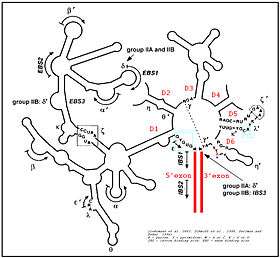
The secondary structure of group II introns is characterized by six typical stem-loop structures, also called domains I to VI or DI to DVI. The domains radiate from a central core that brings the 5' and 3' splice junctions into close proximity. The proximal helix structures of the six domains are connected by a few nucleotides in the central region (linker or joiner sequences). Due to its enormous size, the domain I was divided further into subdomains a, b, c, and d. Sequence differences of group II introns that led to a further division into subgroups IIA, IIB and IIC were identified, along with varying distance of the bulged adenosine in domain VI (the prospective branch point forming the lariat) from the 3' splice site, and the inclusion or omission of structural elements such as a coordination loop in domain I, which is present in IIB and IIC introns but not IIA.[1] Group II introns also form very complicated RNA Tertiary Structure.
Group II introns possess only a very few conserved nucleotides, and the nucleotides important for the catalytic function are spread over the complete intron structure. The few strictly conserved primary sequences are the consensus at the 5' and 3' splicing site (...↓GUGYG&... and ...AY↓..., with the Y representing a pyrimidine), some of the nucleotides of the central core (joiner sequences), a relatively high number of nucleotides of DV and some short-sequence stretches of DI. The unpaired adenosine in DVI (marked by an asterisk in the figure and located 7 or 8 nt away from the 3' splicing site) is also conserved and plays a central role in the splicing process. The 2' hydroxyl of the bulged adenosine attacks the 5' splice site, followed by nucleophilic attack on the 3' splice site by the 3' OH of the upstream exon. This results in a branched intron lariat connected by a 2' phosphodiester linkage at the DVI adenosine.
Protein machinery is required for splicing in vivo, and long-range intron-intron and intron-exon interactions are important for splice site positioning, as well as a number of tertiary contacts between motifs, including kissing-loop and tetraloop-receptor interactions. In 2005, A. De Lencastre et al. found that during splicing of Group II introns, all reactants are preorganized before the initiation of splicing. The branch site, both exons, the catalytically essential regions of DV and J2/3, and ε−ε' are in close proximity before the first step of splicing occurs. In addition to the bulge and AGC triad regions of DV, the J2/3 linker region, the ε−ε' nucleotides and the coordination loop in DI are crucial for the architecture and function of the active-site.[4]
The first crystal structure of a group II intron was resolved in 2008 for the Oceanobacillus iheyensis group IIC catalytic intron, and was joined by the Pylaiella littoralis (P.li.LSUI2) group IIB intron in 2014. Attempts have been made to model the tertiary structure of other group II introns, such as the ai5γ group IIB intron, using a combination of programs for homology mapping onto known structures and de novo modeling of previously unresolved regions.[5]
Distribution and phylogeny
Group II introns are found in rRNA, tRNA, and mRNA of organelles (chloroplasts and mitochondria) in fungi, plants, and protists, and also in mRNA in bacteria. The first intron to be identified as distinct from group I was the ai5γ group IIB intron, which was isolated in 1986 from a pre-mRNA transcript of the oxi 3 mitochondrial gene of Saccharomyces cerevisiae.[6]
A subset of group II introns encode essential splicing proteins, known as intron-encoded proteins or IEPs, in intronic ORFs. The length of these introns can, as a result, be up to 3 kb. Splicing occurs in almost identical fashion to nuclear pre-mRNA splicing with two transesterification steps, with both also using magnesium ions to stabilize the leaving group in each step, which has led some to theorize a phylogenetic link between group II introns and the nuclear spliceosome. Further evidence for this link includes structural similarity between the U2/U6 junction of spliceosomal RNA and domain V of group II introns, which contains the catalytic AGC triad and much of the heart of the active site, as well as parity between conserved 5' and 3' end sequences.[7]
See also
References
- 1 2 Lambowitz AM, Zimmerly S (August 2011). "Group II introns: mobile ribozymes that invade DNA". Cold Spring Harb Perspect Biol. 3 (8): a003616. doi:10.1101/cshperspect.a003616. PMC 3140690. PMID 20463000.
- ↑ Seetharaman, M; Eldho, NV; Padgett, RA; Dayie, KT (Feb 2006). "Structure of a self-splicing group II intron catalytic effector domain 5: parallels with spliceosomal U6 RNA". RNA. 12 (2): 235–47. doi:10.1261/rna.2237806. PMC 1370903. PMID 16428604.
- ↑ Valadkhan, S (May–Jun 2010). "Role of the snRNAs in spliceosomal active site". RNA biology. 7 (3): 345–53. doi:10.4161/rna.7.3.12089. PMID 20458185.
- ↑ de Lencastre A, Hamill S, Pyle AM (July 2005). "A single active-site region for a group II intron". Nat. Struct. Mol. Biol. 12 (7): 626–7. doi:10.1038/nsmb957. PMID 15980867.
- ↑ Somarowthu S, Legiewicz M, Keating KS, Pyle AM (February 2014). "Visualizing the ai5γ group IIB intron". Nucleic Acids Res. 42 (3): 1947–1958. doi:10.1093/nar/gkt1051. PMID 24203709.
- ↑ Peebles CL, Perlman PS, Mecklenburg KL, Petrillo ML, Tabor JH, Jarrell KA, Cheng HL (January 1986). "A self-splicing RNA excises an intron lariat". Cell. 44 (2): 213–223. doi:10.1016/0092-8674(86)90755-5. PMID 3510741.
- ↑ Gordon PM, Sontheimer EJ, Piccirilli JA (February 2000). "Metal ion catalysis during the exon-ligation step of nuclear pre-mRNA splicing: extending the parallels between the spliceosome and group II introns". RNA. 6 (2): 199–205. PMID 10688359.
- Bonen, L; Vogel J (2001). "The ins and outs of group II introns". Trends Genet. 17 (6): 322&ndash, 331. doi:10.1016/S0168-9525(01)02324-1. PMID 11377794.
- Chu, VT; Adamidi C; Liu Q; Perlman PS; Pyle AM (2001). "Control of branch-site choice by a group II intron". EMBO J. 20 (23): 6866&ndash, 6876. doi:10.1093/emboj/20.23.6866. PMC 125754. PMID 11726522.
- Lehmann, K; Schmidt U (2003). "Group II introns: structure and catalytic versatility of large natural ribozymes". Crit Rev Biochem Mol Biol. 38 (3): 249&ndash, 303. doi:10.1080/713609236. PMID 12870716.
- Michel F, Umesono K, Ozeki H (October 1989). "Comparative and functional anatomy of group II catalytic introns--a review". Gene. 82 (1): 5–30. doi:10.1016/0378-1119(89)90026-7. PMID 2684776.