Gluteus medius
| Gluteus medius | |
|---|---|
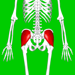 Position of gluteus medius muscle (shown in red). Posterior view. | |
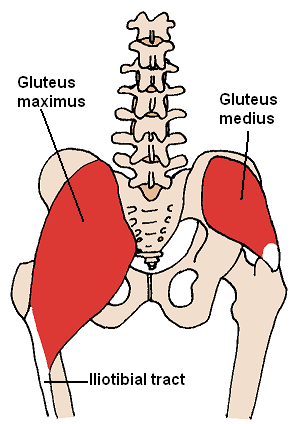 The gluteus medius and nearby muscles. | |
| Details | |
| Origin | Gluteal surface of ilium, under gluteus maximus |
| Insertion | Greater trochanter of the femur |
| Artery | superior gluteal artery |
| Nerve | superior gluteal nerve (L4, L5, S1 nerve roots) |
| Actions | abduction of the hip; preventing adduction of the hip. Medial/internal rotation and flexion of the hip (anterior fibers). Extension and Lateral/external rotation of the hip (posterior fibers) |
| Antagonist | adductors |
| Identifiers | |
| Latin | musculus glutaeus medius |
| TA | A04.7.02.007 |
| FMA | 22315 |
| Anatomical terms of muscle | |
The gluteus medius one of the three gluteal muscles, is a broad, thick, radiating muscle, situated on the outer surface of the pelvis.
Its posterior third is covered by the gluteus maximus, its anterior two-thirds by the gluteal aponeurosis, which separates it from the superficial fascia and integument.
Structure
The gluteus medius muscle starts, or "originates," on the outer surface of the ilium between the iliac crest and the posterior gluteal line above, and the anterior gluteal line below; the gluteus medius also originates from the gluteal aponeurosis that covers its outer surface.
The fibers of the muscle converge into a strong flattened tendon that inserts on the lateral surface of the greater trochanter. More specifically, the muscle's tendon inserts into an oblique ridge that runs downward and forward on the lateral surface of the greater trochanter.
Relations
A bursa separates the tendon of the muscle from the surface of the trochanter over which it glides.
Variations
The posterior border may be more or less closely united to the piriformis, or some of the fibers end on its tendon.
The posterior fibres of gluteus medius contract to produce hip extension, lateral rotation and abduction. During gait, the posterior fibres help to decelerate internal rotation of the femur at the end of swing phase.
Function
• The anterior part acting alone helps to flex and internally rotate the hip.
• The posterior part acting alone helps to extend and externally rotate the hip.
• The anterior and posterior parts working together abduct the hip and stabilizes the pelvis in the coronal plane.[1]
Clinical significance
Dysfunction of the gluteus medius or the superior gluteal nerve can potentially be indicated by a positive Trendelenburg's sign.
Additional images
 Position of gluteus medius muscle (shown in red). Hip bone is shown in semi-transparent.
 Structures surrounding right hip-joint.
 Gluteus medius muscle (shown in green text)
See also
References
This article incorporates text in the public domain from page 474 of the 20th edition of Gray's Anatomy (1918)
- ↑ Schünke, M., Schulte, E., Schumacher, U., Ross, L. M., & Lamperti, E. D. (2006). Thieme atlas of anatomy. Stuttgart: Thieme. page 424
External links
| Wikimedia Commons has media related to Gluteus medius muscles. |
- Anatomy photo:13:st-0404 at the SUNY Downstate Medical Center
- Cross section image: pelvis/pelvis-e12-15—Plastination Laboratory at the Medical University of Vienna