Geniculate ganglion
| Geniculate ganglion | |
|---|---|
 The course and connections of the facial nerve in the temporal bone. | |
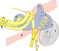 Cranial nerves VII and VIII and selected structures of the inner and middle ear. 1 Nervus vestibularis, 2 Nervus cochlearis, 3 Nervus intermediofacialis, 4 Ganglion geniculi, 5 Chorda tympani, 6 Cochlea, 7 Ductus semicirculares, 8 Malleus, 9 Membrana tympani, 10 Tuba auditiva | |
| Details | |
| Identifiers | |
| Latin | ganglion geniculi nervi facialis |
| MeSH | D005830 |
| TA | A14.2.01.116 |
| FMA | 53414 |
| Anatomical terms of neuroanatomy | |
The geniculate ganglion (from Latin genu, for "knee"[1]) is an L-shaped collection of fibers and pseudo-unipolar sensory neurons of the facial nerve located in the facial canal of the head. It receives fibers from the motor, sensory, and parasympathetic components of the facial nerve and sends fibers that will innervate the lacrimal glands, submandibular glands, sublingual glands, tongue, palate, pharynx, external auditory meatus, stapedius, posterior belly of the digastric muscle, stylohyoid muscle, and muscles of facial expression.
The geniculate ganglion contains special sensory neuronal cell bodies for taste, from fibers coming up from the tongue through the chorda tympani and from fibers coming up from the roof of the palate through the greater petrosal nerve.[2] Sensory and parasympathetic inputs are carried into the geniculate ganglion via the nervus intermedius. Motor fibers are carried via the facial nerve proper. The greater petrosal nerve, which carries preganglionic parasympathetic fibers, emerges from the anterior aspect of the ganglion. The motor fibers of the facial nerve proper and parasympathetic fibers to the submandibular and pterygopalatine ganglia do not synapse in the geniculate ganglion. It is also worth mentioning that the afferent fibers carrying pain, temperature, and touch from the posterior auricular nerve, as well as those carrying special sensory (taste) fibers from the tongue (via the chorda tympani), do not synapse in the geniculate ganglion. Instead, the cells of the geniculate ganglion relay the signal to the appropriate brainstem nucleus, much like the Dorsal root ganglion neurons relay signal to nuclei in the spinal cord[3]
The geniculate ganglion is one of several ganglia of the head and neck. Like the others, it is a bilaterally distributed structure, with each side of the face having a geniculate ganglion.
Additional images
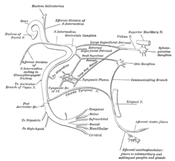 Plan of the facial and intermediate nerves and their communication with other nerves.
See also
References
- ↑ "genu-, geni-, gen- + (Latin: knee)". WordInfo. Retrieved 2008-10-03.
- ↑ M.J. Turlough FitzGerald; Gregory Gruener; Estomih Mtui (2012). Clinical neuroanatomy and neuroscience (6th ed.). Saunders/Elsevier. ISBN 978-0702037382.
- ↑ Moore, Keith L.; Dalley, Arthur F.; Agur, A. M. R. (2013-02-13). Clinically Oriented Anatomy. Lippincott Williams & Wilkins. ISBN 9781451119459.
External links
- cranialnerves at The Anatomy Lesson by Wesley Norman (Georgetown University) (VII)
- lesson3 at The Anatomy Lesson by Wesley Norman (Georgetown University) (midearcavity)