Abdominal external oblique muscle
| Abdominal external oblique muscle | |
|---|---|
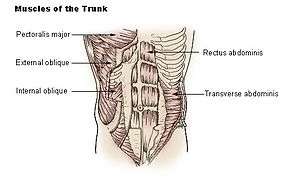 Muscles of the trunk. | |
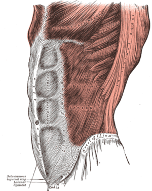 The obliquus externus abdominis | |
| Details | |
| Origin | Ribs 5-12 |
| Insertion | Xiphoid Process, Outer Lip of the Iliac crest, Pubic Crest, Pubic tubercle, Linea alba, Inguinal Ligament, Anterior Superior Iliac Spine (ASIS) |
| Nerve | Thoracoabdominal nerves (T7-11) and Subcostal nerve (T12) |
| Actions | Flexion of the torso and Contralateral rotation of torso |
| Identifiers | |
| Latin | Musculus obliquus externus abdominis |
| TA | A04.5.01.008 |
| FMA | 13335 |
| Anatomical terms of muscle | |
The external oblique muscle (of the abdomen) (also external abdominal oblique muscle) is the largest and the most superficial (outermost) of the three flat muscles of the lateral anterior abdomen.
Structure
The external oblique is situated on the lateral and anterior parts of the abdomen. It is broad, thin, and irregularly quadrilateral, its muscular portion occupying the side, its aponeurosis the anterior wall of the abdomen. In most humans (especially females), the oblique is not visible, due to subcutaneous fat deposits and the small size of the muscle.
It arises from eight fleshy digitations, each from the external surfaces and inferior borders of the fifth to twelfth ribs (lower eight ribs). These digitations are arranged in an oblique line which runs inferiorly and anteriorly, with the upper digitations being attached close to the cartilages of the corresponding ribs, the lowest to the apex of the cartilage of the last rib, the intermediate ones to the ribs at some distance from their cartilages.
The five superior serrations increase in size from above downward, and are received between corresponding processes of the serratus anterior muscle; the three lower ones diminish in size from above downward and receive between them corresponding processes from the latissimus dorsi. From these attachments the fleshy fibers proceed in various directions. Its posterior fibers from the ribs to the iliac crest form a free posterior border.
Those from the lowest ribs pass nearly vertically downward, and are inserted into the anterior half of the outer lip of the iliac crest; the middle and upper fibers, directed downward (inferiorly) and forward (anteriorly), become aponeurotic at approximately the midclavicular line and forms the anterior layer of the rectus sheath. This aponeurosis formed from fibres from either side of the external oblique decussates at the linea alba.
The aponeurosis of the external oblique muscle forms the inguinal ligament. The muscle also contributes to the inguinal canal.
Just deep to the external oblique is the internal oblique muscle. These muscles are in the deepest layer of the abdominal wall.
Nerve supply
The external oblique muscle is supplied by ventral branches of the lower six thoracoabdominal nerves and the subcostal nerve on each side.
Blood supply
The cranial portion of the muscle is supplied by the lower intercostal arteries, whereas the caudal portion is supplied by a branches of either the deep circumflex iliac artery or the iliolumbar artery.
Function
The external oblique functions to pull the chest downwards and compress the abdominal cavity, which increases the intra-abdominal pressure as in a valsalva maneuver. It also performs ipsilateral (same side) side-bending and contralateral (opposite side) rotation. So the right external oblique would side bend to the right and rotate to the left. The internal oblique muscle functions similarly except it rotates ipsilaterally.
Society and culture
Oblique strain
The oblique strain is a common baseball injury, particularly in pitchers. In both batters and pitchers it can affect the contralateral (leading) side external oblique, or the trailing internal oblique.[1]
Training
- Crunch (exercise)
- Side plank
- Sit-up (exercise)
Additional images
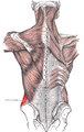 Posterior view of muscles connecting the upper extremity to the vertebral column. Posterior part of abdominal external oblique muscle labeled.
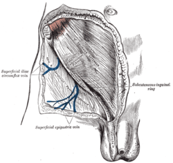 The subcutaneous inguinal ring.
 Transverse section through the middle of the first lumbar vertebra, showing the relations of the pancreas.
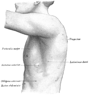 The left side of the thorax.
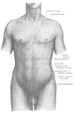 Surface anatomy of the front of the thorax and abdomen.
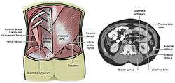 Lumbar triangle
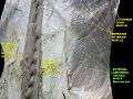 External abdominal oblique muscle. Anterior abdominal wall. Deep dissection. Anterior view.
References
This article incorporates text in the public domain from page 409 of the 20th edition of Gray's Anatomy (1918)
- ↑ Conte, SA; Thompson, MM; Marks, MA; Dines, JS (March 2012). "Abdominal muscle strains in professional baseball: 1991-2010". The American journal of sports medicine. 40 (3): 650–6. doi:10.1177/0363546511433030. PMID 22268233.
External links
- Anatomy image:7061 at the SUNY Downstate Medical Center
- Cross section image: pembody/body8a—Plastination Laboratory at the Medical University of Vienna