Dermatoscopy
| Dermatoscopy | |
|---|---|
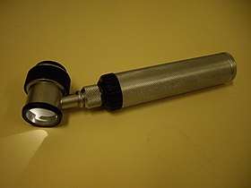 Immersion oil dermatoscope. | |
| MeSH | D046169 |
Dermatoscopy (also known as dermoscopy or epiluminescence microscopy) is the examination of [skin lesions] with a 'dermatoscope'. This traditionally consists of a magnifier (typically x10), a non-polarised light source, a transparent plate and a liquid medium between the instrument and the skin, and allows inspection of skin lesions unobstructed by skin surface reflections. Modern dermatoscopes dispense with the use of liquid medium and instead use polarised light to cancel out skin surface reflections. When the images or video clips are digitally captured or processed, the instrument can be referred to as a "digital epiluminescence dermatoscope".
This instrument is useful to dermatologists in distinguishing benign from malignant (cancerous) lesions, especially in the diagnosis of melanoma.
Types of dermatoscopy
A dermatoscope is composed of a transilluminating light source and a magnifying optic (usually a 10-fold magnification). There are three main modes of dermoscopy:[1]
- Nonpolarized light, contact
- Polarized light, contact
- Polarized light, noncontact
Polarized light allows for visualization of deeper skin structures, while non-polarized light provide information about the superficial skin. Most modern dermatoscopes allow the user to toggle between the two modes, which provide complementary information.
Advantages of dermatoscopy
With doctors who are experts in the specific field of dermoscopy, the diagnostic accuracy for melanoma is significantly better than for those dermatologists who do not have any specialized training in dermatoscopy.[2] Thus, with specialists trained in dermoscopy, there is considerable improvement in the sensitivity (detection of melanomas) as well as specificity (percentage of non-melanomas correctly diagnosed as benign), compared with naked eye examination. The accuracy by dermatoscopy was increased up to 20% in the case of sensitivity and up to 10% in the case of specificity, compared with naked eye examination.[3][4] By using dermatoscopy the specificity is thereby increased, reducing the frequency of unnecessary surgical excisions of benign lesions.[5][6]
In the studies referred to, a comparison is made between dermatoscopy and an artificial situation of only naked eye examination. In reality, most of the 80% of US dermatologists who do not employ dermatoscopy work in bright rooms and use various magnifiers.
Application of dermatoscopy
- The typical application of dermatoscopy is early detection of melanoma (see above)
- Digital dermatoscopy (videodermatoscopy) is used for monitoring skin lesions suspicious of melanoma. Digital dermatoscopy images are stored and compared to images obtained during the patient’s next visit. Suspicious changes in such a lesion are an indication for excision. Skin lesions, which appear unchanged over time are considered benign.[7][8] Common systems for digital dermoscopy are Fotofinder, Molemax, DermoGenius or Easyscan.
- Aid in the diagnosis of skin tumors - such as basal cell carcinomas,[9] squamous cell carcinomas,[10] cylindromas,[11] dermatofibromas, angiomas, seborrheic keratosis and many other common skin tumors have classical dermatoscopic findings.[12]
- Aid in the diagnosis of scabies and pubic louse. By staining the skin with India ink, a dermatoscope can help identify the location of the mite in the burrow, facilitating scraping of the scabetic burrow. By magnifying pubic louse, it allows for rapid diagnosis of the difficult to see small insects.[13][14]
- Aid in the diagnosis of warts. By allowing a physician to visualize the structure of a wart, to distinguish it from corn, callouses, trauma, or foreign bodies. By examining warts at late stages of treatment, to assure that therapy is not stopped prematurely due to difficult to visualize wart structures.
- Aid in the diagnosis of fungal infections. To differentiate "black dot" tinea, or tinea capitis (fungal scalp infection) from alopecia areata.[15]
- Aid in the diagnosis of hair and scalp diseases, such as alopecia areata,[16] female androgenic alopecia,[17] monilethrix,[18] Netherton syndrome,[19] and woolly hair syndrome.[20] Dermoscopy of hair and scalp is called trichoscopy.[21][22]
- Determination of surgical margin of hard to define skin cancers. Examples would be Bowen’s disease, superficial basal cell carcinomas, and lentigo malignas. These tumors have very indistinct margins. By allowing the surgeon to correctly identify the true extent of the tumor, repeat surgery often is decreased.
- Differentiation of tinea nigra from malignant melanoma or junctional melanocytic nevus.[23]
History
Skin surface microscopy started in 1663 by Kolhaus and was improved with the addition of immersion oil in 1878 by Ernst Abbe. The German dermatologist, Johann Saphier, added a built-in light source to the instrument. Goldman was the first dermatologist to coin the term "dermascopy" and to use the dermatoscope to evaluate pigmented cutaneous lesions.
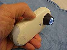
In 2001, a California medical device manufacturer, 3Gen, introduced the first polarized dermatoscope, the DermLite. Polarised illumination, coupled with a cross-polarised viewer, reduces (polarised) skin surface reflection, thus allowing visualisation of skin structures (the light from which is depolarised) without using an immersion fluid. Examination of several lesions is thus more convenient because physicians no longer have to stop and apply immersion oil, alcohol, or water to the skin before examining each lesion. With the marketing of polarised dermatoscopes, dermatoscopy increased in popularity among physicians worldwide. Although images produced by polarised light dermatoscopes are slightly different from those produced by a traditional skin contact glass dermatoscope, they have certain advantages, such as vascular patterns not being potentially missed through compression of the skin by a glass contact plate.
References
- ↑ Argenziano, G; Soyer, HP (July 2001). "Dermoscopy of pigmented skin lesions--a valuable tool for early diagnosis of melanoma". The Lancet. Oncology. 2 (7): 443–9. doi:10.1016/s1470-2045(00)00422-8. PMID 11905739.
- ↑ Lorentzen, H; Weismann, K; Petersen, CS; Larsen, FG; Secher, L; Skødt, V (1999). "Clinical and dermatoscopic diagnosis of malignant melanoma. Assessed by expert and non-expert groups". Acta dermato-venereologica. 79 (4): 301–4. doi:10.1080/000155599750010715. PMID 10429989.
- ↑ Vestergaard, ME; Macaskill, P; Holt, PE; Menzies, SW (2008). "Dermoscopy compared with naked eye examination for the diagnosis of primary melanoma: a meta-analysis of studies performed in a clinical setting". British Journal of Dermatology. 159 (3): 669–76. doi:10.1111/j.1365-2133.2008.08713.x. PMID 18616769. Argenziano, G; Fabbrocini, G; Carli, P; De Giorgi, V; Sammarco, E; Delfino, M (1998). "Epiluminescence microscopy for the diagnosis of doubtful melanocytic skin lesions. Comparison of the ABCD rule of dermatoscopy and a new 7-point checklist based on pattern analysis". Archives of Dermatology. 134 (12): 1563–70. doi:10.1001/archderm.134.12.1563. PMID 9875194.
- ↑ Ascierto, P.A.; Palmieri, G.; Celentano, E.; Parasole, R.; Caraco, C.; Daponte, A.; Chiofalo, M.G.; Melucci, M.T.; Mozzillo, N.; Satriano, R.A.; Castello, G. (2000). "Sensitivity and specificity of epiluminescence microscopy: evaluation on a sample of 2731 excised cutaneous pigmented lesions". British Journal of Dermatology. 142 (5): 893. doi:10.1046/j.1365-2133.2000.03468.x. http://www.bcbstx.com/provider/pdf/medicalpolicies/medicine/201-023.pdf%5Bunreliable+source?%5D
- ↑ Bono, A; Bartoli, C; Cascinelli, N; Lualdi, M; Maurichi, A; Moglia, D; Tragni, G; Tomatis, S; Marchesini, R (2002). "Melanoma detection. A prospective study comparing diagnosis with the naked eye, dermatoscopy and telespectrophotometry". Dermatology. 205 (4): 362–6. doi:10.1159/000066436. PMID 12444332.
- ↑ "Crutchfield Dermatology". Crutchfield Dermatology. Retrieved 2010-05-12.
- ↑ Argenziano, G; Mordente, I; Ferrara, G; Sgambato, A; Annese, P; Zalaudek, I (2008). "Dermoscopic monitoring of melanocytic skin lesions: clinical outcome and patient compliance vary according to follow-up protocols". The British Journal of Dermatology. 159 (2): 331–6. doi:10.1111/j.1365-2133.2008.08649.x. PMID 18510663.
- ↑ Roma, Paolo; Savarese, Imma; Martino, Antonia; Martino, Domenico; Annese, Pietro; Capoluongo, Patrizio; Mordente, Ines; Nicolino, Rachele; Zalaudek, Iris; Argenziano, Giuseppe (2007). "Slow-growing melanoma: Report of five cases". Journal of Dermatological Case Reports. 1. doi:10.3315/jdcr.2007.1.1001. PMC 3157767.
- ↑ Scalvenzi, M; Lembo, S; Francia, MG; Balato, A (2008). "Dermoscopic patterns of superficial basal cell carcinoma". International Journal of Dermatology. 47 (10): 1015–8. doi:10.1111/j.1365-4632.2008.03731.x. PMID 18986346.
- ↑ Felder, S; Rabinovitz, H; Oliviero, M; Kopf, A (2006). "Dermoscopic differentiation of a superficial basal cell carcinoma and squamous cell carcinoma in situ". Dermatologic Surgery. 32 (3): 423–5. doi:10.1111/j.1524-4725.2006.32085.x. PMID 16640692.
- ↑ Sicinska, Justyna; Rakowska, Adriana; Czuwara-Ladykowska, Joanna; Mroz, Andrzej; Lipinski, Marcin; Nasierowska-Guttmejer, Anna; Sikorska, Jolanta; Sklinda, Katarzyna; Slowinska, Monika; Kowalska-Oledzka, Elzbieta; Walecka, Irena; Walecki, Jerzy; Rudnicka, Lidia (2007). "Cylindroma transforming into basal cell carcinoma in a patient with Brooke-Spiegler syndrome". Journal of Dermatological Case Reports. 1. doi:10.3315/jdcr.2007.1.1002. PMC 3157764.
- ↑ Campos-Do-Carmo, G; Ramos-E-Silva, M (2008). "Dermoscopy: basic concepts". International Journal of Dermatology. 47 (7): 712–9. doi:10.1111/j.1365-4632.2008.03556.x. PMID 18613881.
- ↑ Wu, Ming-Yun; Hu, Shu-Lin; Hsu, Che-Hao (June 2008). "Use of Non-contact Dermatoscopy in the Diagnosis of Scabies" (PDF). Dermatol Sinica: 112–4.
- ↑ Chuh, A; Lee, A; Wong, W; Ooi, C; Zawar, V (2007). "Diagnosis of Pediculosis pubis: a novel application of digital epiluminescence dermatoscopy". Journal of the European Academy of Dermatology and Venereology. 21 (6): 837–8. doi:10.1111/j.1468-3083.2006.02040.x. PMID 17567326.
- ↑ Slowinska, M; Rudnicka, L; Schwartz, RA; Kowalska-Oledzka, E; Rakowska, A; Sicinska, J; Lukomska, M; Olszewska, M; Szymanska, E (2008). "Comma hairs: a dermatoscopic marker for tinea capitis: a rapid diagnostic method". Journal of the American Academy of Dermatology. 59 (5 Suppl): S77–9. doi:10.1016/j.jaad.2008.07.009. PMID 19119131.
- ↑ Inui, S; Nakajima, T; Itami, S (2008). "Significance of dermoscopy in acute diffuse and total alopecia of the female scalp: review of twenty cases". Dermatology. 217 (4): 333–6. doi:10.1159/000155644. PMID 18799878.
- ↑ Rakowska, A.; et al. (2008). "Trichoscopy criteria for diagnosing female androgenic alopecia". Nature Precedings. hdl:10101/npre.2008.1913.1.
- ↑ Rakowska, A; Slowinska, M; Czuwara, J; Olszewska, M; Rudnicka, L (2007). "Dermoscopy as a tool for rapid diagnosis of monilethrix". Journal of drugs in dermatology. 6 (2): 222–4. PMID 17373184.
- ↑ Rakowska, A; Kowalska-Oledzka, E; Slowinska, M; Rosinska, D; Rudnicka, L (2009). "Hair shaft videodermoscopy in netherton syndrome". Pediatric dermatology. 26 (3): 320–2. doi:10.1111/j.1525-1470.2008.00778.x. PMID 19706096.
- ↑ Rakowska, Adriana; Slowinska, Monika; Kowalska-Oledzka, Elzbieta; Rudnicka, Lidia (2008). "Trichoscopy in genetic hair shaft abnormalities". Journal of Dermatological Case Reports. 2 (2): 14–20. doi:10.3315/jdcr.2008.1009. PMC 3157768. PMID 21886705.
- ↑ Rudnicka L, Olszewska M, Rakowska A, Kowalska-Oledzka E, Slowinska M (July 2008). "Trichoscopy: a new method for diagnosing hair loss". Journal of Drugs in Dermatology : JDD. 7 (7): 651–4. PMID 18664157.
- ↑ Rakowska A, Slowinska M, Kowalska-Oledzka E, Rudnicka L (2008). "Trichoscopy (hair and scalp videodermoscopy) in the healthy female. Method standardization and norms for measurable parameters". J Dermatol Case Rep. 3 (1): 14–19. doi:10.3315/jdcr.2008.1021. PMC 3157785. PMID 21886722.
- ↑ DOI: https://dx.doi.org/10.1590/abd1806-4841.20142780