Chlamydomonas
| Chlamydomonas | |
|---|---|
.jpg) | |
| SEM image of flagellated Chlamydomonas (10000×) | |
| Scientific classification | |
| (unranked): | Viridiplantae |
| Class: | Chlorophyceae |
| Order: | Chlamydomonadales |
| Family: | Chlamydomonadaceae |
| Genus: | Chlamydomonas Ehrenb. |
| Species | |
|
See text | |
Chlamydomonas is a genus of green algae consisting of about 325 species[1] all unicellular flagellates, found in stagnant water and on damp soil, in freshwater, seawater, and even in snow as "snow algae".[2] Chlamydomonas is used as a model organism for molecular biology, especially studies of flagellar motility and chloroplast dynamics, biogeneses, and genetics. One of the many striking features of Chlamydomonas is that it contains ion channels (channelrhodopsins) that are directly activated by light. Some regulatory systems of Chlamydomonas are more complex than their homologs in Gymnosperms, with evolutionarily related regulatory proteins being larger and containing additional domains.[3]
Molecular phylogeny studies indicated that the traditional genus Chlamydomonas defined using morphological data was polyphyletic within Volvocales, and many species were reclassified (e.g., in Oogamochlamys, Lobochlamys), and many other "Chlamydomonas" lineages are to be reclassificated.[4][5][6]

Description
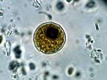
Unicellular cells, spherical or slightly cylindrical with single flagella a papilla may be present or absent. Chloroplasts green and usually cup-shaped.[8]
Species
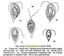
- Chlamydomonas caudata Wille
- Chlamydomonas ehrenbergii Gorozhankin[9]
- Chlamydomonas elegans G.S.West 1915
- Chlamydomonas moewusii
- Chlamydomonas nivalis
- Chlamydomonas ovoidae
- Chlamydomonas reinhardtii[10]
Ecology
Widely distributed in freshwater or damp soil.[11]It is generally found in habitat rich in ammonium salt. Chlamydomonas possesses red eye spots for photosensitivity and reproduces both asexually and sexually.
Chlamydomonas's asexual reproduction occurs by zoospores, by aplanospores, by hypnospores or by a palmella stage;[12] sexual reproduction through isogamy, anisogamy or oogamy.
Nutrition
Most species are obligate phototrophs but C. reinhardtii and C. dysosmos are facultative heterotrophs that can grow in the dark in the presence of acetate as a carbon source.
Morphology
- Motile unicellular algae.
- Generally oval in shape.
- Cell wall is made up of a glycoprotein and non cellulosic polysaccharides instead of cellulose.
- Two anteriorly inserted whiplash flagella. Each flagellum originates from a basal granule in the anterior papillate or non-papillate region of the cytoplasm. Each flagellum shows typical 9+2 arrangement of the component fibrils.
- Contractile vacuoles are near the bases of flagella.
- Prominent cup or bowl shaped chloroplast is present. The chloroplast contains bands composed of a variable number of the photosynthetic thylakoids which are not organised into grana-like structures.
- The nucleus is enclosed in a cup-shaped chloroplast, which has a single large pyrenoid where starch is formed from photosynthetic products. Pyrenoid with starch sheath is present in the posterior end of the chloroplast.
- Eye spot present in the anterior portion of the chloroplast. It consists of two or three, more or less parallel rows of linearly arranged fat droplets.
See also
References
- ↑ Smith, G.M. 1955 Cryptogamic Botany Volume 1. Algae and Fungi McGraw-Hill Book Company Inc
- ↑ Hoham, R.W., Bonome, T.A., Martin, C.W. and Leebens-mack, J.H. 2002. A combined 18S rDNA and rbcL phylogenetic analysis of Chloromonas and Chlamydomonas (Chlorophyceae, Volvocales ) emphasizing snow and other cold-termperature habitats. Journal of Phycology, 38: 1051–1064.
- ↑ A Falciatore, L Merendino, F Barneche, M Ceol, R Meskauskiene, K Apel, JD Rochaix (2005). The FLP proteins act as regulators of chlorophyll synthesis in response to light and plastid signals in Chlamydomonas. The red eye spot in chlamydomonas is sensitive to light and hence determines movement. Genes & Dev, 19:176-187
- ↑ Juliet Brodie & Jane Lewis (2007). Unravelling the algae: the past, present, and future of algal systematics. CRC Press. p. 140, .
- ↑ Wehr, J.D., Sheath, R.G. & Kociolek, J.P. (eds., 2015). Freshwater Algae of North America: Ecology and Classification. Academic Press, USA, p. 275-276, .
- ↑ Pröschold, T., Marin, B., Schlösser, U.W. & Melkonian, M. (2001). Molecular phylogeny and taxonomic revision of Chlamydomonas (Chlorophyta). I. Emendation of Chlamydomonas Ehrenberg and Chloromonas Gobi, and descripription of Oogamochlamys gen. nov. and Lobochlamys gen. nov. Protist 152: 265-300, 7 figs, 5 tables, .
- ↑ Hazen, Tracy E. 1922. The phylogeny of the genus Brachiomonas. Bulletin of the Torrey Botanical Club. 49(4):75-92, with two plates.
- ↑ Guiry, M.D., John, D.M. Rindi, F. and McCarthy, T.K. (ed) 2007 New Survey of Clare Island Volume 6: The Freshwater and Terrestrial Algae. Royal Irish Academy. ISBN 978-1-904890-31-7
- ↑
- ↑ Aoyama, H., Kuroiwa, T and Nakamura,S. 2009. The dynamic behaviour of mitochrandia in living zygotes during maturation and meiosis in Chlamydomonas reinhardtii. European Journal Phycology 44: 497 - 507
- ↑
- ↑ "Life Cycle of Chlamydomonas (With Diagram)". BiologyDiscussion.com. Retrieved 12 March 2018.
External links
- Chlamydomonas Center
- Chlamydomonas reinhardtii Transcription Factor Database
- "Chlamydomonas", a song by Andy Offutt Irwin about the life cycle of Chlamydomonas, posted on the website of the International Society of Protozoologists
- 3D electron microscopy structures of Chlamydomonas-related proteins at the EM Data Bank(EMDB)
- The Seaweed Site