Brain size
The size of the brain is a frequent topic of study within the fields of anatomy and evolution. Brain size can be measured by weight or by volume (via MRI scans or by skull volume). The relationship between brain size and intelligence is frequently a topic of research.
Humans
Studies on human brain size, largely based on participants of European ancestry, tend to find an average adult brain volume of 1260 cubic centimeters (cm3) for men and 1130 cm3 for women. There is, however, substantial variation between individuals;[1] one study of 46 adults, aged 22–49 years and of mainly European descent, found an average brain volume of 1273.6 cm3 for men, with a range of 1052.9 to 1498.5 cm3, and 1131.1 cm3 for women, with a range of 974.9 to 1398.1 cm3.[2]
The right cerebral hemisphere is typically larger than the left, whereas the cerebellar hemispheres are typically closer in size. The adult human brain weighs on average about 1.28 kg (2.8 lb).[3]
Changes over time
Brain size has increased considerably over the course of humans' recent evolutionary history. Homo erectus, a relative of humans, had a brain size of 1,100 cm3. Homo floresiensis, with a brain size of 380 cm3.[4] Neanderthals had a slightly larger brain than modern humans,[5] perhaps due to larger visual systems.[6]
Some studies suggest that the average brain size has been decreasing all over the world over the past 28,000 years, including hunter gatherers like Indigenous Australians. It is possible that the end of the Ice Age had an effect on the diet, muscle mass, or endocrine system of humans, which decreased brain size. [7][8] Others suggest that the cranial capacity for males is unchanged, but that the cranial capacity of females has increased.[9]
Biogeographic variation
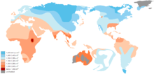
Average cranial capacity in humans varies significantly depending on geographic ancestry in humans, in the range of 1,200 to 1,450 cm3 between populations. Larger cranial volume is associated with climatic region, the largest averages being found in populations of Siberia and the Arctic. For this reason, Beals et al. (1984) proposed that the primary reason for the variation is climatic adaptation, favoring large round heads in colder climates because they conserve heat and slender heads in warm climates closer to the equator (see Bergmann's rule and Allen's rule).[10]
Changes over the lifespan
A baby's brain at birth averages 369 cm3 and increases during the first year of life to about 961 cm3, after which the growth rate declines. Brain volume peaks at the age of 40 years, after which it begins to decline by 5% per decade, speeding up at around 70 years.[11] Total cerebral and gray matter volumes peak between 10 and 20 years (earlier in girls than in boys) of age, whereas white matter and ventricular volumes increase. There is a general pattern in which neural development peaks in childhood and declines in adolescence, a process known as synaptic pruning. Overall white matter volume does not appear to decline with age, although there is variation among brain regions.[12]
Sex
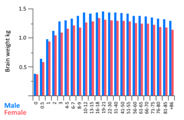
The average brain weight in adult males is 1,345 grams; in adult females, 1,222 grams.[13] Males have been found to have, on average, greater cerebral, cerebellar, and cerebral cortical lobar volumes, except possibly left parietal.[14]
When covaried for intracranial volume, height, and weight, one study found that women have a higher percentage of gray matter, whereas men have a higher percentage of white matter and cerebrospinal fluid. There was high variability among individuals, however.[15] Yaki (2011) found no statistically significant gender differences in the gray matter ratio for most ages in a sample of 758 women and 702 men aged 20–69.[16]
Consistent with findings in adults, average cerebral volume is approximately 10% larger in boys than in girls. However, such differences should not be construed as imparting any sort of functional advantage or disadvantage; gross structural measures may not reflect functionally relevant factors such as neuronal connectivity and receptor density. Moreover, brain volumes, even in narrowly defined groups (e.g. children of the same age), may vary by as much as 50%.[17] Young girls have, on average, larger hippocampi, whereas young boys have larger amygdalas.[15]
Significant dynamic changes in brain structure take place throughout adulthood, with substantial variation between individuals. In later decades, men show greater volume loss in whole brain volume and in the frontal lobes, and temporal lobes, whereas in women there is increased volume loss in the hippocampi and parietal lobes.[15] Men show a steeper decline in global gray matter volume, although in both sexes it varies by region with some areas exhibiting little or no age effect.
Race
In the 19th century, American anthropologist Samuel George Morton reported that whites had the greatest average cranial capacity, followed, in descending order, by Native Americans and blacks. Stephen Jay Gould argued in 1978[18] and in his subsequent book the Mismeasure of Man that Morton unconsciously misrepresented his data and that when it was properly interpreted, it showed no significant racial differences in cranial capacity. This claim was criticized in a 2011 paper, which concluded that Morton did not manipulate his results, unconsciously or otherwise.[19] This paper has, in turn, been criticized for making unwarranted conclusions as to whether Gould's claims of bias were correct or not.[20][21]
J. Philippe Rushton published multiple studies in the 1980s to 2000s claiming that average brain size was lowest in blacks (Negroids) and highest in East Asians (Mongoloids), with whites (Caucasoids) in between the two.[22] His work in this area has been criticized for relying on flawed studies, for failing to consider explanations other than genetics for the observed differences, and for ignoring other studies with contradictory conclusions.[23][24] Nathan Brody has also argued that the evidence regarding racial differences in brain size is not conclusive, and that, even if one accepts it, this difference does not support a genetic hypothesis regarding racial differences in intelligence.[25] Critics of the hereditarian position also note that the difference in mean brain size between blacks and whites is smaller than 1 standard deviation and is insufficient to explain the vast majority of the black-white IQ gap.[26]
Genetic contribution
Adult twin studies indicate that heritability of overall brain size in adulthood is high (between 66% and 97%).[27] Infant brain volumes are also highly heritable, with heritability of total brain volume in neonates of around .8-.9.[28]
Heritability varies regionally within the brain, with high heritabilities of frontal lobe volumes (90-95%), moderate estimates in the hippocampi (40-69%), and environmental factors influencing several medial brain areas. In addition, lateral ventricle volume appears to be mainly explained by environmental factors, suggesting such factors also play a role in the surrounding brain tissue.
Early studies yielded suggestive candidate genes.[27][29] Much larger genome-wide studies have now yielded highly replicable associations for at least 8 genes linked to cortical and subcortical brain volume in a study of over 32,000 humans.[30]
Intelligence
Studies demonstrate a correlation between brain size and intelligence, with larger brains predicting higher intelligence. It is however not clear if the correlation is causal.[31] The majority of MRI studies report moderate correlations around 0.3 to 0.4 between brain volume and intelligence.[32][33] In healthy adults, the correlation of total brain volume and IQ is ~ 0.4 [34] The correlation appears to be related to the known small correlation of height with intelligence, which can be explained almost entirely by greater brain volume.[35]
Studies have sought particular regions that are more correlated with IQ than whole-brain volume. While consistent associations are observed within the frontal, temporal, and parietal lobes, the hippocampus, and the cerebellum, unique variation in these regions account for a relatively small amount of variance in IQ.[33][36] The search for specific brain regions that correlate highly with cognitive measures designed to be specific has not yielded clear results.[2]
There may be sex differences in the volume-IQ association. Some evidence suggests that while IQ correlates equally with frontal lobe volume, it may correlate more with parietal lobe volumes in men and with Broca's area in women, corresponding to spatial versus language specializations.[15]
Small studies have attempted to link brain volume with functional measures such as P300 auditory evoked potentials but finding no association.[37][38] Studies attempting to related sibling differences in IQ to differences in brain volume are hampered by relatively small sample sizes and the noisy nature of such difference scores, yielding weak evidence for cross-trait cross-sib correlations.[31]
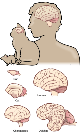
Other animals
The largest brains are those of sperm whales, weighing about 8 kg (18 lb), and killer whales, weighing about 12–15 lb (5.4–6.8 kg). An elephant's brain weighs just over 5 kg (11 lb) and a bottlenose dolphin's 1.5 to 1.7 kg (3.3 to 3.7 lb).
Brain size tends to vary according to body size. The relationship is not proportional, however; the brain-to-body mass ratio varies. The largest ratio found is in the shrew.[39] Averaging brain weight across all orders of mammals, it follows a power law, with an exponent of about 0.75.[40] This power law formula applies to the "average" brain of mammals taken as a whole, but each family (cats, rodents, primates, etc.) departs from it to some degree, in a way that generally reflects the overall "sophistication" of behavior.[41] Primates, for a given body size, have brains 5 to 10 times as large as the formula predicts. Predators tend to have relatively larger brains than the animals they prey on; placental mammals (the great majority) have relatively larger brains than marsupials such as the opossum.
When the mammalian brain increases in size, not all parts increase at the same rate.[42] In particular, the larger the brain of a species, the greater the fraction taken up by the cortex. Thus, in the species with the largest brains, most of their volume is filled with cortex: this applies not only to humans, but also to animals such as dolphins, whales or elephants.
Not all investigators are happy with the amount of attention that has been paid to brain size. Roth and Dicke, for example, have argued that factors other than size are more highly correlated with intelligence, such as the number of cortical neurons and the speed of their connections.[43] Moreover, they point out that intelligence depends not just on the amount of brain tissue, but on the details of how it is structured. It is also well known that crows, ravens, and grey parrots are quite intelligent even though they have small brains.
Cranial capacity
Cranial capacity is a measure of the volume of the interior of the cranium (also called the braincase or brainpan or skull) of those vertebrates who have both a cranium and a brain. The volume of the cranium is used as a rough indicator of the size of the brain, although due to the thickness of the membranes that surround the brain, brain volume is less than cranial capacity. Cranial Capacity is often tested by filling the cranial cavity with particulate material (as mustard seed or small shot) and measuring the volume of the latter. However, this method of measuring cranial capacity must be validated in each species to know whether it is an accurate representation of the braincase.[44][45]
Knowledge of the volume of the cranial cavity can be important information for the study of different populations with various differences like geographical, racial, or ethnic origin. Other things, such as nutrition, can also affect cranial capacity.[46]
The volume of the human braincase has increased as humans have evolved (see Homininae), starting from about 600 cm3 in Homo habilis up to 1740 cm3 in Homo neanderthalensis, which was the hominin with the biggest brain size.[47][48]
In an attempt to use cranial capacity as an objective indicator of brain size, the encephalization quotient (EQ) was developed in 1973 by Harry Jerison. It compares the size of the brain of the specimen to the expected brain size of animals with roughly the same weight.[49] This way a more objective judgement can be made on the cranial capacity of an individual animal. A large scientific collection of brain endocasts and measurements of cranial capacity has been compiled by Holloway et al. (2005).[50]
Examples of cranial capacity
Apes
- Orangutans: 275–500 cm3 (16.8–30.5 cu in)
- Chimpanzees: 275–500 cm3 (16.8–30.5 cu in)
- Gorillas: 340–752 cm3 (20.7–45.9 cu in)
Hominins
- Australopithecus afarensis: 445.8 cm3 (27.20 cu in)
- Australopithecus africanus: 491.2 cm3 (29.97 cu in)
- Homo habilis: 610.3 cm3 (37.24 cu in)
- Homo erectus: 1,092.9 cm3 (66.69 cu in)
- Homo erectus soloensis: 1,155.8 cm3 (70.53 cu in)
- Homo heidelbergensis: 1,262.8 cm3 (77.06 cu in)
- Homo neanderthalensis: 1,427.2 cm3 (87.09 cu in)
- Homo sapiens: 1,496.5 cm3 (91.32 cu in)
- Individual range, without cephalic disorder: 900–2,100 cm3 (55–128 cu in)
- Global group means range: 1,085–1,581 cm3 (66.2–96.5 cu in)
- Average: 1,353 cm3 (82.6 cu in)
See also
Notes
- ↑ Cosgrove, KP; Mazure CM; Staley JK (2007). "Evolving knowledge of sex differences in brain structure, function, and chemistry". Biol Psychiatry. 62 (8): 847–55. doi:10.1016/j.biopsych.2007.03.001. PMC 2711771. PMID 17544382.
- 1 2 Allen et al., 2002
- ↑ Kelley Hays; David S. Reader in Gender archaeology. Routlegde. Retrieved 2014-09-21.
- ↑ Brown, P.; Sutikna, T.; Morwood, M. J.; Soejono, R. P.; Jatmiko; Saptomo, E. Wayhu; Due, Rokus Awe (October 2004). "A new small-bodied hominin from the Late Pleistocene of Flores, Indonesia". Nature. 431 (7012): 1055–1061. doi:10.1038/nature02999. PMID 15514638.
- ↑ Holloway, 1995
- ↑ Pearce, Eiluned; Stringer, Chris; Dunbar, R. I. M. (2013-05-07). "New insights into differences in brain organization between Neanderthals and anatomically modern humans". Proceedings of the Royal Society of London B: Biological Sciences. 280 (1758): 20130168. doi:10.1098/rspb.2013.0168. ISSN 0962-8452. PMC 3619466. PMID 23486442.
- ↑ "If Modern Humans Are So Smart, Why Are Our Brains Shrinking?". DiscoverMagazine.com. 2011-01-20. Retrieved 2014-03-05.
- ↑ Henneberg, Maciej (1988). "Decrease of human skull size in the Holocene". Human Biology. 60: 395–405. JSTOR 41464021.
- ↑ Rushton, John Philippe (1992). "Cranial capacity related to sex, rank, and race in a stratified random sample of 6,325 U.S. military personnel". Intelligence. 16 (3–4): 401–413. doi:10.1016/0160-2896(92)90017-l.
- 1 2 Kenneth L. Beals, Courtland L. Smith, and Stephen M. Dodd, "Brain Size, Cranial Morphology, Climate, and Time Machines" CURRENT ANTHROPOLOGY V01. 25, NO 01984 (3 June 1984), fig. p. 304. "We offer an alternative hypothesis that suggests that hominid expansion into regions of cold climate produced change in head shape. Such change in shape contributed to the increased cranial volume. Bioclimatic effects directly upon body size (and indirectly upon brain size) in combination with cranial globularity appear to be a fairly powerful explanation of ethnic group differences." "Morphological Adaptation to Climate in Modern Homo sapiens Crania: The Importance of Basicranial Breadth". Wioletta Nowaczewska, Pawe D browski1 and Lukasz KuŸmiñski.
- ↑ Peters, R. (2006). "Ageing and the brain". Postgraduate Medical Journal. 82: 84–8. doi:10.1136/pgmj.2005.036665. PMC 2596698. PMID 16461469.
- ↑ Good et al., 2001
- ↑ Kelley Hays; David S. Reader in Gender archaeology. Routlegde. Retrieved 2014-09-21.
- ↑ Carne et al., 2006
- 1 2 3 4 Cosgrove et al., 2007
- ↑ Taki, Y.; Thyreau, B.; Kinomura, S.; Sato, K.; Goto, R.; Kawashima, R.; Fukuda, H. (2011). He, Yong, ed. "Correlations among Brain Gray Matter Volumes, Age, Gender, and Hemisphere in Healthy Individuals". PLoS ONE. 6 (7): e22734. doi:10.1371/journal.pone.0022734. PMC 3144937. PMID 21818377.
- ↑ Giedd, 2008
- ↑ Gould, S. J. (1978-05-05). "Morton's ranking of races by cranial capacity. Unconscious manipulation of data may be a scientific norm". Science. 200 (4341): 503–509. doi:10.1126/science.347573. ISSN 0036-8075. PMID 347573.
- ↑ Lewis, Jason E.; DeGusta, David; Meyer, Marc R.; Monge, Janet M.; Mann, Alan E.; Holloway, Ralph L. (2011-06-07). "The Mismeasure of Science: Stephen Jay Gould versus Samuel George Morton on Skulls and Bias". PLOS Biology. 9 (6): e1001071. doi:10.1371/journal.pbio.1001071. ISSN 1545-7885.
- ↑ Weisberg, Michael (May 2014). "Remeasuring man". Evolution & Development. 16 (3): 166–178. doi:10.1111/ede.12077. ISSN 1525-142X. PMID 24761929.
- ↑ Weisberg, Michael; Paul, Diane B. (2016-04-19). "Morton, Gould, and Bias: A Comment on "The Mismeasure of Science"". PLOS Biology. 14 (4): e1002444. doi:10.1371/journal.pbio.1002444. ISSN 1545-7885.
- ↑ Rushton, J.Philippe (January 1988). "Race differences in behaviour: A review and evolutionary analysis". Personality and Individual Differences. 9 (6): 1009–1024. doi:10.1016/0191-8869(88)90135-3. . Rushton, J. Philippe; Ankney, C. Davison (March 1996). "Brain size and cognitive ability: Correlations with age, sex, social class, and race". Psychonomic Bulletin & Review. 3 (1): 21–36. doi:10.3758/BF03210739. . Rushton, J. Philippe; Rushton, Elizabeth W. (March–April 2003). "Brain size, IQ, and racial-group differences: Evidence from musculoskeletal traits". Intelligence. 31 (2): 139–155. doi:10.1016/S0160-2896(02)00137-X. .
- ↑ Cain, Donald P.; Vanderwolf, C.H. (1990). "A critique of Rushton on race, brain size and intelligence". Personality and Individual Differences. 11 (8): 777–784. doi:10.1016/0191-8869(90)90185-T.
- ↑ Kamin, Leon J.; Omari, Safiya (1998-09-01). "Race, Head Size, and Intelligence". South African Journal of Psychology. 28 (3): 119–128. doi:10.1177/008124639802800301. ISSN 0081-2463.
We describe errors in published reports, and find that American whites have greater head height than American blacks, but that blacks have greater head length and greater head circumference.
- ↑ Brody, Nathan (2003). "Jensen's Genetic Interpretation of Racial Differences in Intelligence: Critical Evaluation". The Scientific Study of General Intelligence: Tribute to Arthur Jensen. Elsevier Science. pp. 397–410.
- ↑ Wicherts, Jelte M.; Borsboom, Denny; Dolan, Conor V. (January 2010). "Evolution, brain size, and the national IQ of peoples around 3000 years B.C". Personality and Individual Differences. 48 (2): 104–106. doi:10.1016/j.paid.2009.08.020.
- 1 2 Peper, 2007
- ↑ Gilmore, John H.; Schmitt, James Eric; Knickmeyer, Rebecca C.; Smith, Jeffrey K.; Lin, Weili; Styner, Martin; Gerig, Guido; Neale, Michael C. (2010-08-01). "Genetic and environmental contributions to neonatal brain structure: A twin study". Human Brain Mapping. 31 (8): 1174–1182. doi:10.1002/hbm.20926. ISSN 1097-0193. PMC 3109622.
- ↑ Zhang, 2003
- ↑ Adams, H.H. (2016). "Novel genetic loci underlying human intracranial volume identified through genome-wide association". Nature Neuroscience. 19: 1569–1582. doi:10.1038/nn.4398.
- 1 2 Nisbett et al. 2012b, p. 142.
- ↑ McDaniel, Michael (2005). "Big-brained people are smarter" (PDF). Intelligence. 33: 337–346. doi:10.1016/j.intell.2004.11.005.
- 1 2 Luders et al., 2008
- ↑ Gignac, Gilles E.; Bates, Timothy C. (2017). "Brain volume and intelligence: The moderating role of intelligence measurement quality". Intelligence. 64: 18–29. doi:10.1016/j.intell.2017.06.004.
- ↑ Vuoksimaa; et al. (2017). "Brain structure mediates the association between height and cognitive ability". bioRxiv 183525.
- ↑ Hoppe & Stojanovic, 2008
- ↑ Egan et al., 1993
- ↑ Egan et al, 1995
- ↑ Kevin Kelly. "The Technium: Brains of White Matter". kk.org.
- ↑ Armstrong, 1983
- ↑ Jerison, Evolution of the Brain and Intelligence
- ↑ Finlay et al., 2001
- ↑ Roth & Dicke, 2005
- ↑ Logan & Clutton-Brock (2013). "Validating methods for estimating endocranial volume in individual red deer (Cervus elaphus)" (PDF). Behavioural Processes. 92: 143–146. doi:10.1016/j.beproc.2012.10.015.
- ↑ Logan & Palmstrom (2015). "Can endocranial volume be estimated accurately from external skull measurements in great-tailed grackles (Quiscalus mexicanus)?". PeerJ. 3: e1000. doi:10.7717/peerj.1000. PMC 4465945. PMID 26082858.
- ↑ J. Philippe Rushton; Arthur R. Jensen. "THIRTY YEARS OF RESEARCH ON RACE DIFFERENCES IN COGNITIVE ABILITY" (PDF). American Psychological Association.
- ↑ Amano, Hideki (2015). "Virtual reconstruction of the Neanderthal Amud 1 cranium". American Journal of Physical Anthropology. 158: 185–197. doi:10.1002/ajpa.22777.
- ↑ Brown, Graham; Fairfax, Stephanie; Sarao, Nidhi. "Human Evolution". Tree of Life. Tree of Life Project. Retrieved 19 May 2016.
- ↑ Campbell, G.C., Loy, J.D., Cruz-Uribe, K. (2006). Humankind Emerging: Ninth Edition. Boston: Pearson. p346
- ↑ Holloway, Ralph L., Yuan, M. S., and Broadfield, D.C. (2005). The Human Fossil Record: Brain Endocasts: The Paleoneurological Evidence. New York. John Wiley & Sons Publishers (http://www.columbia.edu/~rlh2/PartII.pdf and http://www.columbia.edu/~rlh2/available_pdfs.htmlfor further references). Archived 2014-06-01 at the Wayback Machine.
- ↑ Smith, C. L., Beals, K. L. (1990). "Cultural correlates with cranial capacity". American Anthropologist. 92: 193–200. doi:10.1525/aa.1990.92.1.02a00150.
- ↑ Beals, Kenneth L.; Smith, Courtland L.; Dodd, Stephen M. (June 1984). "Brain Size, Cranial Morphology, Climate, and Time Machines" (PDF). Current Anthropology. 25 (3): 301–330. doi:10.1086/203138. Retrieved 28 January 2018.
References
- Aiello, L; Wheeler, P (1995). "The Expensive Tissue Hypothesis: The Brain and the Digestive System in Human and Primate Evolution" (PDF). Current Anthropology. 36 (2): 199–221. doi:10.1086/204350. Retrieved 15 April 2011.
- Allen, JS; Damasio H; Grabowski TJ (2002). "Normal neuroanatomical variation in the human brain: An MRI-volumetric study". Am J Phys Anthropol. 118 (4): 341–58. doi:10.1002/ajpa.10092. PMID 12124914.
- Armstrong, E (1983). "Relative brain size and metabolism in mammals". Science. 220 (4603): 1302–4. doi:10.1126/science.6407108. PMID 6407108.
- Carne, RP; Vogrin S; Litewka L; Cook MJ (2006). "Cerebral cortex: An MRI-based study of volume and variance with age and sex". J Clin Neurosci. 13 (1): 60–72. doi:10.1016/j.jocn.2005.02.013. PMID 16410199.
- Cosgrove, KP; Mazure CM; Staley JK (2007). "Evolving Knowledge of Sex Differences in Brain Structure, Function and Chemistry". Biol Psychiatry. 62 (8): 847–55. doi:10.1016/j.biopsych.2007.03.001. PMC 2711771. PMID 17544382.
- Driemeyer, J; Boyke, J; Gaser, C; Buchel, C; May, A (2008). Eagleman, David M., ed. "Changes in Gray Matter Induced by Learning—Revisited". PLoS ONE. 3 (7): 7. doi:10.1371/journal.pone.0002669. PMC 2447176. PMID 18648501.

- Egan, V; Chiswick A; Santosh C; Naidu K; Rimmington JE; Best JJK (1993). "Size isn't everything: A study of brain volume, intelligence and auditory evoked potentials". Pers Ind Diff. 17 (3): 357–367. doi:10.1016/0191-8869(94)90283-6.
- Egan, V; Wickett JC; Vernon PA (1995). "Brain size and intelligence: Erratum, addendum, and correction" (PDF). Personality and Individual Differences. 19 (1): 113–115. doi:10.1016/0191-8869(95)00043-6.
- Finlay, BL; Darlington RB; Nicastro N (2001). "Developmental structure in brain evolution" (PDF). Behav Brain Sci. 24 (2): 263–308. doi:10.1017/S0140525X01003958. PMID 11530543. Archived from the original (PDF) on 2008-10-31. Retrieved 2008-11-01.
- Giedd, JN (2008). "The teen brain: insights from neuroimaging" (PDF). J Adolescent Health. 42 (4): 335–43. doi:10.1016/j.jadohealth.2008.01.007. PMID 18346658. Archived from the original (PDF) on 2008-10-31. Retrieved 2008-11-01.
- Good CD, Johnsrude IS, Ashburner J, Henson RN, Friston KJ, Frackowiak RS (2001). "A voxel-based morphometric study of ageing in 465 normal adult human brains". NeuroImage. 14 (1 Pt 1): 21–36. doi:10.1006/nimg.2001.0786. PMID 11525331.
- Holloway, RL (1995). Changeaux JP, Chavillon J, ed. Origins of the Human Brain. Clarendon. pp. 42–54. ISBN 978-0-19-852307-9.
- Ilg, R; Wohlschläger AM; Gaser C; Liebau Y; Dauner R; Wöller A; Zimmer C; Zihl J; Mühlau M (2008). "Gray matter increase induced by practice correlates with task-specific activation: a combined functional and morphometric magnetic resonance imaging study". J Neurosci. 28 (16): 4210–5. doi:10.1523/JNEUROSCI.5722-07.2008. PMID 18417700.
- Jerison, HJ (1973). Evolution of the Brain and Intelligence. Academic Press. ISBN 978-0-12-385250-2.
- Kappelman, J (1993). "The evolution of body mass and relative brain size in fossil hominids". Journal of Human Evolution. 30 (3): 243–76. doi:10.1006/jhev.1996.0021.
- Lange, N; Giedd JN; Castellanos FX; Vaituzis AC; Rapoport JL (1997). "Variability of human brain structure size: ages 4–20 years". Psychiat Res: Neuroimaging. 74 (6): 1–12. doi:10.1016/S0925-4927(96)03054-5. PMID 10710158.
- Lee, H; Devlin JT; Shakeshaft C; Stewart LH; Brennan A; Glensman J; Pitcher K; Crinion J; Mechelli A; Frackowiak RS; Green DW; Price CJ (2007). "Anatomical traces of vocabulary acquisition in the adolescent brain". J Neurosci. 27 (5): 1184–9. doi:10.1523/JNEUROSCI.4442-06.2007. PMID 17267574.
- Hoppe, C; Stojanovic J (2008). "High-aptitude minds: the neurological roots of genius". Scientific American.
- Luders, E; Narr KL; Thompson PM; Toga AW (2008). "Neuroanatomical Correlates of Intelligence". Intelligence. 37 (2): 156–163. doi:10.1016/j.intell.2008.07.002. PMC 2770698. PMID 20160919.
- Peper, JS; Brouwer, RM; Boomsma, DI; Kahn, RS; Hulshoff Pol, HE (2007). "Genetic influences on human brain structure: A review of brain imaging studies in twins". Human Brain Mapping. 28 (6): 464–73. doi:10.1002/hbm.20398. PMID 17415783.
- Ross, C; Henneberg M (1995). "Basicranial flexion, relative brain size, and facial kyphosis in Homo sapiens and some fossil hominids" (PDF). Am J Phys Anthropol. 98 (4): 575–93. doi:10.1002/ajpa.1330980413. PMID 8599387. Archived from the original (PDF) on 2010-06-12. Retrieved 2008-11-01.
- Roth, G; Dicke U (2005). "Evolution of the brain and intelligence" (PDF). Trends Cogn Sci. 9 (5): 250–7. doi:10.1016/j.tics.2005.03.005. PMID 15866152.
- Savage, MV; Gillooly JF; Woodruff WH; West GB; Allen AP; Enquist BJ; Brown JH (2004). "The predominance of quarter-power scaling in biology". Functional Ecol. 18 (2): 257–82. doi:10.1111/j.0269-8463.2004.00856.x.
- Zhang, J (2003). "Evolution of the human ASPM gene, a major determinant of brain size". Genetics. 165 (4): 2063–70. PMC 1462882. PMID 14704186.
Further reading
- Jabr, Ferris (28 November 2015). "How Humans Ended Up With Freakishly Huge Brains". Wired. Retrieved 29 November 2015.
| Cognition | |
|---|---|
| Intelligence | |
| Pain | |
| Relation to brain | |
| |