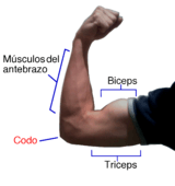
Biceps
| Biceps | |
|---|---|
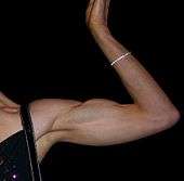
 The biceps is a two-headed muscle and is one of the chief flexors of the forearm. Here is the left side, seen from the front. | |
| Details | |
| Pronunciation |
/ˈbaɪsɛps |
| Origin |
Short head: coracoid process of the scapula. Long head: supraglenoid tubercle |
| Insertion | Radial tuberosity and bicipital aponeurosis into deep fascia on medial part of forearm |
| Artery | Brachial artery |
| Nerve | Musculocutaneous nerve (C5–C7)[1] |
| Actions | |
| Antagonist | Triceps brachii muscle |
| Identifiers | |
| Latin | musculus biceps brachii |
| TA | A04.6.02.013 |
| FMA | 37670 |
| Anatomical terms of muscle | |
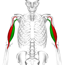
The biceps, also biceps brachii (Latin for "two-headed muscle of the arm"), is a large muscle that lies on the front of the upper arm between the shoulder and the elbow. Both heads of the muscle arise on the scapula and join to form a single muscle belly which is attached to the upper forearm. While the biceps crosses both the shoulder and elbow joints, its main function is at the elbow where it flexes the forearm and supinates the forearm. Both these movements are used when opening a bottle with a corkscrew: first biceps unscrews the cork (supination), then it pulls the cork out (flexion).[2]
Structure
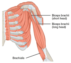
The biceps is one of three muscles in the anterior compartment of the upper arm, along with the brachialis muscle and the coracobrachialis muscle, with which the biceps shares a nerve supply.[1] The biceps muscle has two heads, the short head and the long head, distinguished according to their origin at the coracoid process and supraglenoid tubercle of the scapula, respectively.[1] From its origin on the glenoid, the long head remains tendinous as it passes through the shoulder joint and through the intertubercular groove of the humerus.[2] Extending from its origin on the coracoid, the tendon of the short head runs adjacent to the tendon of the coracobrachialis as the conjoint tendon. Unlike the other muscles in the anterior compartment of the arm, the biceps muscle crosses two joints, the shoulder joint and the elbow joint.
Both heads of the biceps join in the middle upper arm to form a single muscle mass usually near the insertion of the deltoid to form a common muscle belly, although several anatomic studies have demonstrated that the muscle bellies remain distinct structures without confluent fibers.[3][4] As the muscle extends distally, the two heads rotate 90 degrees externally before inserting onto the radial tuberosity. The short head inserts distally on the tuberosity while the long head inserts proximally closer to the apex of the tuberosity.[3] The bicipital aponeurosis, also called the lacertus fibrosus, is a thick fascial band that organizes close to the musculotendinous junction of the biceps and radiates over and inserts onto the ulnar part of the antebrachial fascia.[5]
The tendon that attaches to the radial tuberosity is partially or completely surrounded by a bursa, the bicipitoradial bursa, which ensures frictionless motion between the biceps tendon and the proximal radius during pronation and supination of the forearm.[6]
Two muscles lie underneath the biceps brachii. These are the coracobrachialis muscle, which like the biceps attaches to the coracoid process of the scapula, and the brachialis muscle which connects to the ulna and along the mid-shaft of the humerus. Besides those, the brachioradialis muscle is adjacent to the biceps and also inserts on the radius bone, though more distally.
Variation
Traditionally described as a two-headed muscle, biceps brachii is one of the most variable muscles of the human body and has a third head arising from the humerus in 10% of cases (normal variation)—most commonly originating near the insertion of the coracobrachialis and joining the short head—but four, five, and even seven supernumerary heads have been reported in rare cases.[7]
The distal biceps tendons are completely separated in 40% and bifurcated in 25% of cases. [8][4]
Nerve supply
The biceps shares its nerve supply with the other two muscles of the anterior compartment. The muscles are supplied by the musculocutaneous nerve. Fibers of the fifth, sixth and seventh cervical nerves make up the components of the musculocutaneous nerve which supply the biceps.[1]
Function

The biceps works across three joints.[9] The most important of these functions is to supinate the forearm and flex the elbow. Besides, the long head of biceps prevents the upward displacement of the head of the humerus.[10] In more detail, the actions are, by joint:[11]
- Proximal radioulnar joint (upper forearm) – Contrary to popular belief, the biceps brachii is not the most powerful flexor of the forearm, a role which actually belongs to the deeper brachialis muscle. The biceps brachii functions primarily as a powerful supinator of the forearm (turns the palm upwards). This action, which is aided by the supinator muscle, requires the elbow to be at least partially flexed. If the elbow, or humeroulnar joint, is fully extended, supination is then primarily carried out by the supinator muscle. The biceps is a particularly powerful supinator of the forearm due to the distal attachment of the muscle at the radial tuberosity, on the opposite side of the bone from the supinator muscle. When flexed, the biceps effectively pulls the radius back into its neutral supinated position in concert with the supinator muscle.[12]:346–347
- Elbow (Humeroulnar joint) – The biceps brachii also functions as an important flexor of the forearm, particularly when the forearm is supinated.[1] Functionally, this action is performed when lifting an object, such as a bag of groceries or when performing a biceps curl. When the forearm is in pronation (the palm faces the ground), the brachialis, brachioradialis, and supinator function to flex the forearm, with minimal contribution from the biceps brachii. It is also important to note that regardless of forearm position, (supinated, pronated, or neutral,) the force exerted by the biceps brachii remains the same; however, the brachioradialis has a much greater change in exertion depending on position than the biceps during concentric contractions. That is, the biceps can only exert so much force, and as forearm position changes, other muscles must compensate.[13]
- Shoulder (Glenohumeral joint) (shoulder) – Several weaker functions occur at the glenohumeral, or shoulder, joint. The biceps brachii weakly assists in forward flexion of the shoulder joint (bringing the arm forward and upwards). It may also contribute to abduction (bringing the arm out to the side) when the arm is externally (or laterally) rotated. The short head of the biceps brachii also assists with horizontal adduction (bringing the arm across the body) when the arm is internally (or medially) rotated. Finally, the short head of the biceps brachii, due to its attachment to the scapula (or shoulder blade), assists with stabilization of the shoulder joint when a heavy weight is carried in the arm. The tendon of the long head of the biceps also assists in holding the head of the humerus in the glenoid cavity.[12]:295
Clinical significance
The proximal tendons of the biceps brachii are commonly involved in pathological processes and are a frequent cause of anterior shoulder pain.[14] Disorders of the distal biceps brachii tendon include insertional tendonitis and partial or complete tears of the tendon. Partial tears are usually characterized by pain and enlargement and abnormal contour of the tendon.[15] Complete tears occur as avulsion of the tendinous portion of the biceps away from its insertion on the tuberosity of the radius, and is often accompanied by a palpable, audible "pop" and immediate pain and soft tissue swelling.[16] A soft-tissue mass is sometimes encountered in the anterior aspect of the arm, the so-called Reverse Popeye deformity, which paradoxically leads to a decreased strength during flexion of the elbow and supination of the forearm.[17] Tears of the biceps brachii may occur during athletic activities, however avulsion injuries of the distal biceps tendon are frequently occupational in nature and sustained during forceful, eccentric contraction of the biceps muscle while lifting.[16] Acute rupture of the distal biceps tendon can be treated nonoperatively with acceptable results,[18] but because the injury can lead to 30% loss of elbow flexion strength and 30-50% loss of forearm supination strength, surgical repair is generally recommended.[19][20][21] Complete distal biceps tears are commonly treated with re-attachment of the biceps tendon to its native insertion on the tuberosity of the radius using bone tunnels, suture buttons, or suture anchors.[22][19][23] Proximal ruptures of the long head of the biceps tendon can be surgically addressed by two different techniques. Biceps tenodesis includes release of the long head of the biceps tendon off of its insertion on the glenoid and re-attachment by screw or suture anchor fixation to the humerus. Biceps tenotomy consists of simple release of the long head of the biceps without reattachment to the humerus, allowing the tendon to retract into the soft tissues of the proximal upper arm.[16] Degeneration of the tendon can cause partial tears and are rarely associated with a traumatic event. Treatment of a biceps tear depends on the severity of the injury. In most cases, the muscle will heal over time with no corrective surgery. Applying cold pressure and using anti-inflammatory medications will ease pain and reduce swelling. More severe injuries require surgery and post-op physical therapy to regain strength and functionality in the muscle. Corrective surgeries of this nature are typically reserved for elite athletes who rely on a complete recovery.[24]
Society and culture
The biceps are usually attributed as representative of strength within a variety of worldwide cultures.
Etymology and grammar
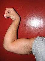
The biceps brachii muscle is the one that gave all muscles their name: it comes from the Latin musculus, "little mouse", because the appearance of the flexed biceps resembles the back of a mouse. The same phenomenon occurred in Greek, in which μῦς, mȳs, means both "mouse" and "muscle".
The term biceps brachii is a Latin phrase meaning "two-headed [muscle] of the arm", in reference to the fact that the muscle consists of two bundles of muscle, each with its own origin, sharing a common insertion point near the elbow joint. The proper plural form of the Latin adjective biceps is bicipites, a form not in general English use. Instead, biceps is used in both singular and plural (i.e., when referring to both arms).
The English form bicep [sic], attested from 1939, is a back formation derived from interpreting the s of biceps as the English plural marker -s.[25][26] While common even in professional contexts, it is often considered incorrect.[27]
Training
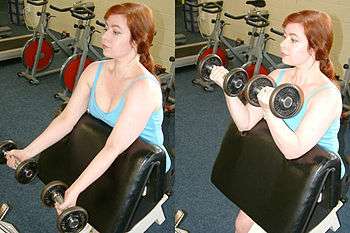
The biceps can be strengthened using weight and resistance training. Examples of well known biceps exercises are the chin-up and biceps curl.
To isolate the biceps brachii in elbow flexion, place the shoulder in hyperextension.
In training the biceps brachii, it is important to distinguish between the long head and the short head of the biceps. The long head is the outer portion of the muscle. This portion originates in the shoulder joint and is a common source of shoulder pain.[28] The short head is the inner portion of the muscle. This portion originates on the coracoid process.[29]
There is much debate over the best biceps workouts for targeting each of these heads.
The first theory for targeting is based on the proximity of the arms in relation to the body. It is said that when the elbows are pulled back behind the body, this targets the long head more. To target the short head, the elbows should be in front of the body.
The second theory uses grip placement and angle as the primary factor in targeting each head. For instance, to target the long head when using dumbbells or cables, the grip should be semi-supinated (hammer) grip where the palms face each other. If using a barbell (EZ grip or straight), the grip should be inside of shoulder width. To target the short head when using dumbbells or cables, grip should be supinated, where the palms are facing up completely. If using a barbell (EZ grip or straight), grip should be outside of shoulder width.[30]
History
Leonardo da Vinci expressed the original idea of the biceps acting as a supinator in a series of annotated drawings made between 1505 and 1510; in which the principle of the biceps as a supinator, as well as its role as a flexor to the elbow were devised. However, this function remained undiscovered by the medical community as da Vinci was not regarded as a teacher of anatomy, nor were his results publicly released. It was not until 1713 that this movement was re-discovered by William Cheselden and subsequently recorded for the medical community. It was rewritten several times by different authors wishing to present information to different audiences. The most notable recent expansion upon Cheselden's recordings was written by Guillaume Duchenne in 1867, in a journal named Physiology of Motion. To this day it remains one of the major references on supination action of the biceps brachii.
Other species
Neanderthals
In Neanderthals, the radial bicipital tuberosities were larger than in modern humans, which suggests they were probably able to use their biceps for supination over a wider range of pronation-supination. It is possible that they relied more on their biceps for forceful supination without the assistance of the supinator muscle like in modern humans, and thus that they used a different movement when throwing.[31]
Horses
In the horse, the biceps' function is to extend the shoulder and flex the elbow. It is composed of two short-fibred heads separated longitudinally by a thick internal tendon which stretches from the origin on the supraglenoid tubercle to the insertion on the medial radial tuberosity. This tendon can withstand very large forces when the biceps is stretched. From this internal tendon a strip of tendon, the lacertus fibrosus, connects the muscle with the extensor carpi radialis -- an important feature in the horse's stay apparatus (through which the horse can rest and sleep whilst standing.) [32]
Additional images
 Biceps and triceps.
 Movement of biceps and triceps when arm is flexing
References
- 1 2 3 4 5 6 7 8 Bogart BI, Ort VH (2007). Elsevier's integrated anatomy and embryology. Philadelphia, Pa.: Elsevier Saunders. pp. 262–267. ISBN 978-1-4160-3165-9.
- 1 2 Lippert LS (2006). Clinical kinesiology and anatomy (4th ed.). Philadelphia: F. A. Davis Company. pp. 126–7. ISBN 978-0-8036-1243-3.
- 1 2 Athwal GS, Steinmann SP, Rispoli DM (October 2007). "The distal biceps tendon: footprint and relevant clinical anatomy". The Journal of Hand Surgery. 32 (8): 1225–9. doi:10.1016/j.jhsa.2007.05.027. PMID 17923307.
- 1 2 Eames MH, Bain GI, Fogg QA, van Riet RP (May 2007). "Distal biceps tendon anatomy: a cadaveric study". The Journal of Bone and Joint Surgery. American Volume. 89 (5): 1044–9. doi:10.2106/JBJS.D.02992. PMID 17473142.
- ↑ Platzer W (2004). Color Atlas of Human Anatomy. Vol. 1: Locomotor System (5th ed.). Thieme. p. 154. ISBN 1-58890-159-9.
- ↑ Kegels L, Van Oyen J, Siemons W, Verdonk R (June 2006). "Bicipitoradial bursitis. A case report" (PDF). Acta Orthopaedica Belgica. 72 (3): 362–5. PMID 16889153.
- ↑ Poudel PP, Bhattarai C (June 2009). "Study on the supernumerary heads of biceps brachii muscle in Nepalese" (PDF). Nepal Medical College Journal. 11 (2): 96–8. PMID 19968147.
- ↑ Dirim B, Brouha SS, Pretterklieber ML, Wolff KS, Frank A, Pathria MN, Chung CB (December 2008). "Terminal bifurcation of the biceps brachii muscle and tendon: anatomic considerations and clinical implications". AJR. American Journal of Roentgenology. 191 (6): W248–55. doi:10.2214/AJR.08.1048. PMID 19020211.
- ↑ "Biceps Brachii". ExRx.net. Retrieved 16 January 2017.
- ↑ Krishna G (2010). "8 - Arm". BD Chaurasia's Human Anatomy (Regional and Applied Dissection and Clinical) Volume 1 - Upper limb and thorax (Fifth ed.). India: CBS Publishers and Distributors Pvt Ltd. p. 88. ISBN 978-81-239-1863-1.
- ↑ Simons DG, Travell JG, Simons LS (1999). "30: Biceps Brachii Muscle". In Eric Johnson. Travell & Simons' Myofascial Pain and Dysfunction (2nd ed.). Baltimore, Maryland: Williams and Wilkins. pp. 648–659. ISBN 0-683-08363-5.
- 1 2 Saladin K (2015). Anatomy and Physiology: The Unity of Form and Function. New York, NY: McGraw-Hill Education. ISBN 978-0-07-340371-7.
- ↑ Kleiber T, Kunz L, Disselhorst-Klug C (2015-01-01). "Muscular coordination of biceps brachii and brachioradialis in elbow flexion with respect to hand position". Frontiers in Physiology. 6: 215. doi:10.3389/fphys.2015.00215. PMC 4526813. PMID 26300781.
- ↑ Frost A, Zafar MS, Maffulli N (April 2009). "Tenotomy versus tenodesis in the management of pathologic lesions of the tendon of the long head of the biceps brachii". The American Journal of Sports Medicine. 37 (4): 828–33. doi:10.1177/0363546508322179. PMID 18762669.
- ↑ Chew ML, Giuffrè BM (2005). "Disorders of the distal biceps brachii tendon". Radiographics. 25 (5): 1227–37. doi:10.1148/rg.255045160. PMID 16160108.
- 1 2 3 Miller MD, Thompson SR, DeLee J, Drez D (2015). DeLee & Drez's orthopaedic sports medicine : principles and practice (Fourth ed.). Philadelphia, PA. ISBN 978-1-4557-4376-6. OCLC 880421005.
- ↑ Arend CF. Ultrasound of the Shoulder. Master Medical Books, 2013. Free chapter on ultrasound evaluation of biceps tendon tears available at ShoulderUS.com
- ↑ Freeman CR, McCormick KR, Mahoney D, Baratz M, Lubahn JD (October 2009). "Nonoperative treatment of distal biceps tendon ruptures compared with a historical control group". The Journal of Bone and Joint Surgery. American Volume. 91 (10): 2329–34. doi:10.2106/jbjs.h.01150. PMID 19797566.
- 1 2 Morrey BF, Askew LJ, An KN, Dobyns JH (March 1985). "Rupture of the distal tendon of the biceps brachii. A biomechanical study". The Journal of Bone and Joint Surgery. American Volume. 67 (3): 418–21. PMID 3972866.
- ↑ Baker BE, Bierwagen D (March 1985). "Rupture of the distal tendon of the biceps brachii. Operative versus non-operative treatment". The Journal of Bone and Joint Surgery. American Volume. 67 (3): 414–7. PMID 3972865.
- ↑ Nesterenko S, Domire ZJ, Morrey BF, Sanchez-Sotelo J (March 2010). "Elbow strength and endurance in patients with a ruptured distal biceps tendon". Journal of Shoulder and Elbow Surgery. 19 (2): 184–9. doi:10.1016/j.jse.2009.06.001. PMID 19664936.
- ↑ Sotereanos DG, Pierce TD, Varitimidis SE (May 2000). "A simplified method for repair of distal biceps tendon ruptures". Journal of Shoulder and Elbow Surgery. 9 (3): 227–33. PMID 10888168.
- ↑ Bain GI, Prem H, Heptinstall RJ, Verhellen R, Paix D (March 2000). "Repair of distal biceps tendon rupture: a new technique using the Endobutton". Journal of Shoulder and Elbow Surgery. 9 (2): 120–6. PMID 10810691.
- ↑ "Bicep tear - Muscular Injuries". Sports Medicine Information.
- ↑ "Bicep". Dictionary and Thesaurus — Merriam-Webster Online. Retrieved December 22, 2010.
- ↑ Zwicky A (July 30, 2008). "The dangers of satire". Language Log. Retrieved December 22, 2010.
- ↑ Corbett PB (February 9, 2010). "Tangled Passages". The New York Times.
- ↑ AlQahtani SM, Bicknell RT (December 2016). "Outcomes following long head of biceps tendon tenodesis". Current Reviews in Musculoskeletal Medicine. 9 (4): 378–387. doi:10.1007/s12178-016-9362-7. PMC 5127942. PMID 27599831.
- ↑ Mohammed H, Skalski MR, Patel DB, Tomasian A, Schein AJ, White EA, Hatch GF, Matcuk GR (2016). "Coracoid Process: The Lighthouse of the Shoulder". Radiographics. 36 (7): 2084–2101. doi:10.1148/rg.2016160039. PMID 27471875.
- ↑ "Biceps Workouts".
- ↑ Churchill SE, Rhodes JA (2009). "The Evolution of the Human Capacity for 'Killing at a Distance': The Human Fossil Evidence for the Evolution of Projectile Weaponry". In Hublin JJ, Richards MP. The evolution of hominin diets: integrating approaches to the study of Palaeolithic subsistence. Springer Science + Business Media. p. 208. ISBN 978-1-4020-9698-3.
- ↑ Watson JC, Wilson AM (January 2007). "Muscle architecture of biceps brachii, triceps brachii and supraspinatus in the horse". Journal of Anatomy. 210 (1): 32–40. doi:10.1111/j.1469-7580.2006.00669.x. PMC 2100266. PMID 17229281.
External links
| Wikimedia Commons has media related to Biceps brachii muscle. |
| Look up biceps in Wiktionary, the free dictionary. |
- Anatomy photo:06:05-0102 at the SUNY Downstate Medical Center
- "Bicep Exercises". Elite Men's Guide Health.