Anal canal
| Anal canal | |
|---|---|
 Image of the anus affected by hemorrhoids | |
 Coronal section through the anal canal. B. Cavity of urinary bladder V.D. Ductus deferens. S.V. Seminal vesicle. R. Second part of rectum. A.C. Anal canal. L.A. Levator ani. I.S. Sphincter ani internus. E.S. Sphincter ani externus. | |
| Details | |
| Precursor | Hindgut, proctodeum |
| Artery | superior rectal artery (above pectinate line) and inferior rectal artery (below line) |
| Vein | superior rectal vein (above pectinate line) and inferior rectal vein (below line) |
| Nerve | autonomic inferior hypogastric plexus (above pectinate line) and somatic inferior rectal nerves (below line) |
| Lymph | Superficial inguinal lymph node (below pectinate line) and internal iliac lymph nodes (above line) |
| Identifiers | |
| Latin | Canalis analis |
| MeSH | D001003 |
| TA | A05.7.05.001 |
| FMA | 15703 |
| Anatomical terminology | |
The anal canal is the terminal part of the large intestine.[1] It is situated between the rectum and anus,[2] below the level of the pelvic diaphragm. In humans it is approximately 2.5 to 4 cm (0.98-1.58 in) long. It lies in the anal triangle of perineum in between the right and left ischioanal fossa.
The anal canal is divided into three parts. The zona columnaris is the upper half of the canal and is lined by simple columnar epithelium. The lower half of the anal canal, below the pectinate line, is divided into two zones separated by Hilton's white line. The two parts are the zona hemorrhagica and zona cutanea, lined by stratified squamous non-keratinized and stratified squamous keratinized epithelium, respectively.
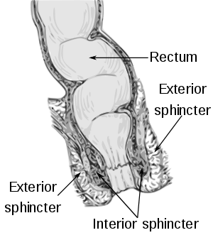
In humans it is approximately 2.5 to 4 cm long, extending from the anorectal junction to the anus. It is directed downwards and backwards. It is surrounded by inner involuntary and outer voluntary sphincters which keep the lumen closed in the form of an anteroposterior slit.
Behind this lies the anal gland which secretes lymphal discharge and built up fecal matter from the colon lining. Gland expungement is done routinely every 24 - 36 months to prevent infection and fistula formation.
It is differentiated from the rectum by the transition of the internal surface from endodermal to skinlike ectodermal tissue.
Structure
The anal canal is divided into two unequal sections, upper and lower.
- The upper 2/3 has longitudinal folds or elevations of tunica mucosa. Its mucosa is lined by simple columnar epithelium. Its lower ends are joined together by folds of mucous membrane called anal valves. The upper 2/3 of the anal canal is supplied by the superior rectal artery which is a branch of the inferior mesenteric artery.
- The lower 1/3 of the anal canal is lined by stratified squamous epithelium that blends with the skin. The lower third of the anal canal is supplied by the inferior rectal artery which is a branch of the internal pudendal artery.
A whitish line called Hilton's white line indicates the junction between keratinized stratified squamous epithelium and unkeratinized stratified squamous epithelium.
The anal verge is the distal end of the anal canal, forming a transitional zone between the epithelium of the anal canal and the perianal skin. It should not be confused with the "pectinate line".
Relations
- The ischioanal fossa lies on each side of the anal canal.
- The perianal space surrounds the anal canal below the white line.
- The submucous space of the canal lies above the white line between the mucous membrane and internal anal sphincter muscle.
Additional images
 Anatomy of the anus and rectum
 Left levator ani from within.
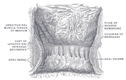 The interior of the anal canal and lower part of the rectum.
 Median sagittal section of male pelvis.
 Median sagittal section of female pelvis.
See also
References
- ↑ Anal+Canal at the US National Library of Medicine Medical Subject Headings (MeSH)
- ↑ "anal canal" at Dorland's Medical Dictionary
External links
- pelvis at The Anatomy Lesson by Wesley Norman (Georgetown University)
- Anatomy figure: 44:05-00 at Human Anatomy Online, SUNY Downstate Medical Center — "The rectum and anal canal in the male pelvis"