Actomyosin ring
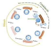
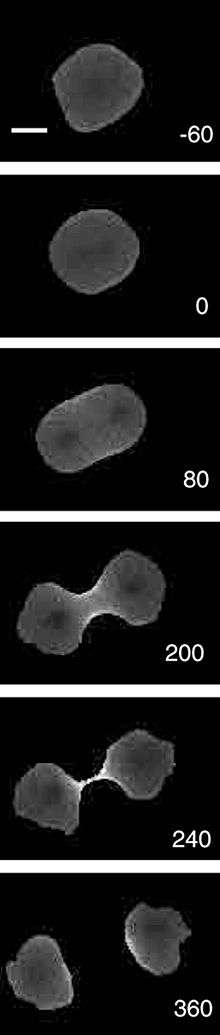
The cell cycle is divided into two primary phases: DNA synthesis or S phase and Mitosis or M phase. During the S phase, duplication of chromosomes occurs whereas M phase is characterized by two processes known as nuclear (mitosis) and cytoplasmic (cytokinesis) divisions. The Actomyosin ring is a prominent structure during cytoplasmic division or cytokinesis[1] (i.e., splitting from the parent cell after duplication and segregation of the genetic material has been completed). The ring forms perpendicular to the axis of the spindle apparatus.[2] and occurs towards the later stage of mitosis, the telophase, in which sister chromatids are identically separated at the opposite sides of the spindle forming nuclei (see figure 1). The actomyosin ring follows an orderly sequence of identification of active division site, formation of the ring, constriction of the ring, and disassembly of the contractile ring.[1] It is composed of actin and myosin II bundles, thus the term actomyosin, that operates in contractile motion [3] although the mechanism on how or what triggers the constriction is still an evolving topic.[4] Other cytoskeletal proteins are also involved in maintaining stability of the ring.[5] Apart from cytokinesis in which the ring constricts as the cells divide (see figure 2), actomyosin ring constriction has also been found to activate during wound closure.[6] During the process actin filaments are degraded, preserving the thickness of the ring. After cytokinesis is complete, one of the two daughter cells inherits a remnant, called the midbody ring.[7]
Activation of the cell-cycle kinase (e.g. Rho-kinases) during telophase initiates constriction of the actomyosin ring by creating a groove that migrates in an inward motion. Rho-kinases such as ROCK1 has been found to regulate actomyosin contraction through phosphorylation of the myosin light chain (MLC).[8] This mechanism promotes cell-cell contacts and integrity leading to adhesion formation.
Variation between Kingdoms
In animals, the ring forms along the cleavage furrow on the inside of the plasma membrane then splits by abscission.[9][10] In fungi it forms at the mother-bud neck before mitosis. Septin is heavily involved in the formation of the fungal AMR.[11] In most bacteria and many archea a homologous structure called the z-ring forms out of FtsZ, a homolog of tubulin.[12] Chloroplasts form an analogous structure out of FtsZ. These structures are not made out of actomyosin but serve a similar role in constricting and permitting cytokinesis. In plant cells, there is no actomyosin ring. Instead a cell plate grows centrifugally outwards from the center of the plane of division until it fuses with the existing cell wall.[13]
See also
References
- 1 2 Cheffings TH, Burroughs NJ, Balasubramanian MK. (2016). Actomyosin Ring Formation and Tension Generation in Eukaryotic Cytokinesis. Curr Biol. 26(15):719-737.
- ↑ Mana-Capelli, S. & McCollum, D. (2013). Actomyosin Ring. Encyclopedia of Systems Biology 8-8. doi:10.1007/978-1-4419-9863-7_779 URL: https://link.springer.com/referenceworkentry/10.1007%2F978-1-4419-9863-7_779
- ↑ Munjal, Akankshi and Lecuit, Thomas. (2014). Actomyosin networks and tissue morphogenesis. Development. 141(9):1789-1793.
- ↑ Mendes Pinto, I., Rubinstein, B., & Li, R. (2013). Force to Divide: Structural and Mechanical Requirements for Actomyosin Ring Contraction. Biophysical Journal. 105(3):547–554.
- ↑ Martin, Adam. (2016). Embryonic ring closure: Actomyosin rings do the two-step. J. Cell. Biol. 215(3):301-303.
- ↑ Cornelia Schwayer, Mateusz Sikora, Jana Slováková, Roland Kardos, Carl-Philipp Heisenberg. (2016). Actin Rings of Power. In: Developmental Cell. 37(6): 493-506.
- ↑ Pelletier, L., & Yamashita, Y. (2012). Centrosome asymmetry and inheritance during animal development. Current Opinion In Cell Biology. 24(4): 541-546. https://dx.doi.org/10.1016/j.ceb.2012.05.005
- ↑ Shi, J., Surma, M., Zhang, L., & Wei, L. (2013). Dissecting the roles of ROCK isoforms in stress-induced cell detachment. Cell Cycle. 12(10): 1492–1500.
- ↑ Chen, Chun-ting, H. Hehnly, and S. Doxsey. (2012). Orchestrating vesicle transport, ESCRTs and kinase surveillance during abscission. Nature reviews. Molecular Cell Biology. 13: 483-488.
- ↑ Fededa, J. P., and D.W. Gerlich. (2012). Molecular control of animal cell cytokinesis. Nature Cell Biology, 14(5): 440-447.
- ↑ Meitinger, F. and S. Palani. (2016). Actomyosin ring driven cytokinesis in budding yeast. Seminars in Cell & Developmental Biology 53:19-27.
- ↑ Alberts, B., A. Johnson, J. Lewis, D. Morgan, M. Raff, K. Roberts, and P. Walter, editors. (2015). Molecular Biology of the Cell, 6th edition. Garland Science: New York. 1464 pp.
- ↑ Bi, E., P. Maddox, D. Lew, E. Salmon, J. McMillan, E. Yeh, and J. Pringle. (1998). Involvement of an Actomyosin Contractile Ring in Saccharomyces cerevisiae Cytokinesis. Journal Of Cell Biology. 142(5):1301–1312.