Accessory navicular bone
| Accessory navicular bone | |
|---|---|
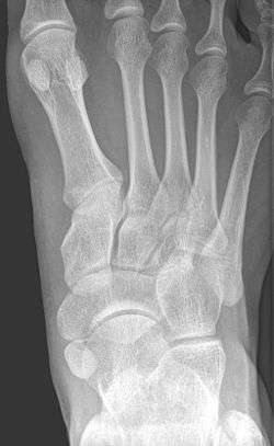 X-ray of the foot showing an accessory navicular bone | |
| Details | |
| Identifiers | |
| Latin | Os tibiale, os tibiale externum or naviculare secundarium |
| MeSH | C536002 |
| Anatomical terms of bone | |
An accessory navicular bone is an accessory bone of the foot that occasionally develops abnormally in front of the ankle towards the inside of the foot. This bone may be present in approximately 2-21% of the general population and is usually asymptomatic.[1][2][3] When it is symptomatic, surgery may be necessary.
Accessory navicular bone may cause a continuous stretch and stress on the tibialis posterior tendon which can progress to chronic disabling pain and may cause tendon rupture or secondary flat foot deformity, when this occurs this condition is commonly known as accessory navicular syndrome.[4]
Other conditions which closely mimic the symptoms of an accessory navicular bone include plantar fasciitis, bunions and heel spurs.
Classification
The Geist classification divides the accessory navicular bones into three types.[3]
- Type 1: An os tibiale externum is a 2–3 mm sesamoid bone in the distal posterior tibialis tendon. Usually asymptomatic.
- Type 2: Triangular or heart-shaped ossicle measuring up to 12 mm, which represents a secondary ossification center connected to the navicular tuberosity by a 1–2 mm layer of fibrocartilage or hyaline cartilage. Portions of the posterior tibialis tendon sometimes insert onto the accessory ossicle, which can cause dysfunction, and therefore, symptoms.
- Type 3: A cornuate navicular bone represents an enlarged navicular tuberosity, which may represent a fused Type 2 accessory bone. Occasionally symptomatic due to bunion formation.
Treatment
Aside from surgery, there are a few options for handling an accessory navicular bone that has become symptomatic. This includes immobilization, icing, medicating, physical therapy, and orthotic devices. Immobilizing involves placing the foot and ankle in a cast or removable walking boot. This alleviates stressors on the foot and can decrease inflammation. Icing will help reduce swelling and inflammation. Medication involves usage of nonsteroidal anti-inflammatory drugs, or steroids (taken orally or injected) to decrease inflammation. Physical therapy can be prescribed in order to strengthen the muscles and help decrease inflammation. Physical therapy can also help prevent the symptoms from returning. Orthotic devices (arch support devices that fit in a shoe) can help prevent future symptoms.[5] Occasionally, the orthotic device will dig into the edge of the accessory navicular and cause discomfort. For this reason, the orthotic devices made for the patient should be carefully constructed.[6]
Gross images
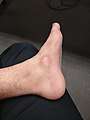 Accessory navicular bone prominence.
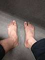 Foot with accessory navicular (left) compared with a normal foot (right).
 Flat foot deformity caused by accessory navicular bone.
Radiological images
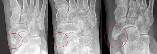 From left to right: Type 1, 2 and 3
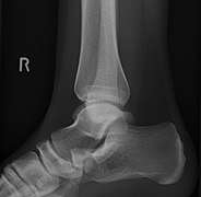 Lateral projection of type 2
 Type 2
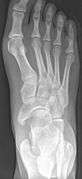 Cornuate navicular bone
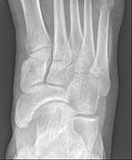 Cornuate navicular bone
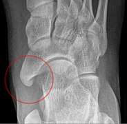 Cornuate navicular bone
 Type 2 on one foot (dark arrow) and type 3 on the other (white arrow)
References
- ↑ http://www.wheelessonline.com/ortho/accessory_navicular
- ↑ http://www.macrorad.com/case-reports/symptomatic-accessory-navicular-bone.html
- 1 2 Miller TT, Staron RB, Feldman F, Parisien M, Glucksman WJ, Gandolfo LH (June 1995). "The symptomatic accessory tarsal navicular bone: assessment with MR imaging". Radiology. 195 (3): 849–853. doi:10.1148/radiology.195.3.7754020. PMID 7754020.
- ↑ Sahibzada N. Mansoora, Farooq A.Rathore (October 2007). "Symptomatic accessory navicular bone: A case series". The Egyptian Rheumatologist. 39 (4): 263–266. doi:10.1016/j.ejr.2017.02.003.
- ↑ http://www.foothealthfacts.org/footankleinfo/accessory_navicular_syndrome.htm
- ↑ http://mdmercy.com/centers-of-excellence/orthopedics-bone-and-joint/institute-for-foot-and-ankle-reconstruction/conditions-we-treat/flat-feet/accessory-navicular?sc_lang=en
- Golano, P; Fariñas, O; Sáenz, I (March 2004). "The anatomy of the navicular and periarticular structures". Foot and ankle clinics. 9 (1): 1–23. doi:10.1016/S1083-7515(03)00155-4. PMID 15062212.
External links

- Wheeless' Textbook of Orthopaedics
- Mercy Institute for foot and ankle reconstruction
- Montana spine center
- Macrorad Teleradiology