ATPIF1

ATPase inhibitor, mitochondrial is an enzyme that in humans is encoded by the ATPIF1 gene.[5][6]
This gene encodes a mitochondrial ATPase inhibitor. Alternative splicing occurs at this locus and three transcript variants encoding distinct isoforms have been identified.[6]
It prevents ATPase from switching to ATP hydrolysis during collapse of the electrochemical gradient, for example during oxygen deprivation [7] ATP synthase inhibitor forms a one-to-one complex with the F1 ATPase, possibly by binding at the alpha-beta interface. It is thought to inhibit ATP synthesis by preventing the release of ATP.[8] The inhibitor has two oligomeric states, dimer (the active state) and tetramer. At low pH, the inhibitor forms a dimer via antiparallel coiled coil interactions between the C-terminal regions of two monomers. At high pH, the inhibitor forms tetramers and higher oligomers by coiled coil interactions involving the N terminus and inhibitory region, thus preventing the inhibitory activity.[7]
Model organisms
| Characteristic | Phenotype |
|---|---|
| Homozygote viability | Normal |
| Fertility | Normal |
| Body weight | Normal |
| Anxiety | Abnormal[9] |
| Neurological assessment | Normal |
| Grip strength | Normal |
| Hot plate | Normal |
| Dysmorphology | Normal |
| Indirect calorimetry | Normal |
| Glucose tolerance test | Normal |
| Auditory brainstem response | Normal |
| DEXA | Normal |
| Radiography | Normal |
| Body temperature | Normal |
| Eye morphology | Normal |
| Clinical chemistry | Abnormal[10] |
| Haematology | Normal |
| Peripheral blood lymphocytes | Normal |
| Micronucleus test | Normal |
| Heart weight | Normal |
| Brain histopathology | Abnormal |
| Salmonella infection | Normal[11] |
| Citrobacter infection | Normal[12] |
| All tests and analysis from[13][14] |
Model organisms have been used in the study of ATPIF1 function. A conditional knockout mouse line, called Atpif1tm1a(EUCOMM)Wtsi[15][16] was generated as part of the International Knockout Mouse Consortium program — a high-throughput mutagenesis project to generate and distribute animal models of disease to interested scientists.[17][18][19]
Male and female animals underwent a standardized phenotypic screen to determine the effects of deletion.[13][20] Twenty three tests were carried out on mutant mice and three significant abnormalities were observed.[13] Homozygous mutant animals displayed hyperactivity and brain dysmorphology, while males also had decreased circulating alkaline phosphatase levels.[13]
| Mitochondrial ATPase inhibitor, IATP | |||||||||
|---|---|---|---|---|---|---|---|---|---|
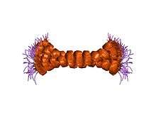 c-terminal coiled-coil domain from bovine if1 | |||||||||
| Identifiers | |||||||||
| Symbol | IATP | ||||||||
| Pfam | PF04568 | ||||||||
| InterPro | IPR007648 | ||||||||
| SCOP | 1hf9 | ||||||||
| SUPERFAMILY | 1hf9 | ||||||||
| |||||||||
References
- 1 2 3 ENSG00000285390 GRCh38: Ensembl release 89: ENSG00000130770, ENSG00000285390 - Ensembl, May 2017
- 1 2 3 GRCm38: Ensembl release 89: ENSMUSG00000054428 - Ensembl, May 2017
- ↑ "Human PubMed Reference:".
- ↑ "Mouse PubMed Reference:".
- ↑ Ichikawa N, Ushida S, Kawabata M, Masazumi Y (Mar 2000). "Nucleotide sequence of cDNA coding the mitochondrial precursor protein of the ATPase inhibitor from humans". Biosci Biotechnol Biochem. 63 (12): 2225–2227. doi:10.1271/bbb.63.2225. PMID 10664857.
- 1 2 "Entrez Gene: ATPIF1 ATPase inhibitory factor 1".
- 1 2 Cabezon E, Butler PJ, Runswick MJ, Carbajo RJ, Walker JE (November 2002). "Homologous and heterologous inhibitory effects of ATPase inhibitor proteins on F-ATPases". J. Biol. Chem. 277 (44): 41334–41. doi:10.1074/jbc.M207169200. PMID 12186878.
- ↑ van Raaij MJ, Orriss GL, Montgomery MG, Runswick MJ, Fearnley IM, Skehel JM, Walker JE (December 1996). "The ATPase inhibitor protein from bovine heart mitochondria: the minimal inhibitory sequence". Biochemistry. 35 (49): 15618–25. doi:10.1021/bi960628f. PMID 8961923.
- ↑ "Anxiety data for Atpif1". Wellcome Trust Sanger Institute.
- ↑ "Clinical chemistry data for Atpif1". Wellcome Trust Sanger Institute.
- ↑ "Salmonella infection data for Atpif1". Wellcome Trust Sanger Institute.
- ↑ "Citrobacter infection data for Atpif1". Wellcome Trust Sanger Institute.
- 1 2 3 4 Gerdin AK (2010). "The Sanger Mouse Genetics Programme: High throughput characterisation of knockout mice". Acta Ophthalmologica. 88: 925–7. doi:10.1111/j.1755-3768.2010.4142.x.
- ↑ Mouse Resources Portal, Wellcome Trust Sanger Institute.
- ↑ "International Knockout Mouse Consortium".
- ↑ "Mouse Genome Informatics".
- ↑ Skarnes, W. C.; Rosen, B.; West, A. P.; Koutsourakis, M.; Bushell, W.; Iyer, V.; Mujica, A. O.; Thomas, M.; Harrow, J.; Cox, T.; Jackson, D.; Severin, J.; Biggs, P.; Fu, J.; Nefedov, M.; De Jong, P. J.; Stewart, A. F.; Bradley, A. (2011). "A conditional knockout resource for the genome-wide study of mouse gene function". Nature. 474 (7351): 337–342. doi:10.1038/nature10163. PMC 3572410. PMID 21677750.
- ↑ Dolgin E (2011). "Mouse library set to be knockout". Nature. 474 (7351): 262–3. doi:10.1038/474262a. PMID 21677718.
- ↑ Collins FS, Rossant J, Wurst W (2007). "A Mouse for All Reasons". Cell. 128 (1): 9–13. doi:10.1016/j.cell.2006.12.018. PMID 17218247.
- ↑ van der Weyden L, White JK, Adams DJ, Logan DW (2011). "The mouse genetics toolkit: revealing function and mechanism". Genome Biol. 12 (6): 224. doi:10.1186/gb-2011-12-6-224. PMC 3218837. PMID 21722353.
External links
- Human ATPIF1 genome location and ATPIF1 gene details page in the UCSC Genome Browser.
Further reading
- Cabezón E, Runswick MJ, Leslie AG, Walker JE (2002). "The structure of bovine IF(1), the regulatory subunit of mitochondrial F-ATPase". EMBO J. 20 (24): 6990–6996. doi:10.1093/emboj/20.24.6990. PMC 125800. PMID 11742976.
- Strausberg RL, Feingold EA, Grouse LH, et al. (2003). "Generation and initial analysis of more than 15,000 full-length human and mouse cDNA sequences". Proc. Natl. Acad. Sci. U.S.A. 99 (26): 16899–16903. doi:10.1073/pnas.242603899. PMC 139241. PMID 12477932.
- Ota T, Suzuki Y, Nishikawa T, et al. (2004). "Complete sequencing and characterization of 21,243 full-length human cDNAs". Nat. Genet. 36 (1): 40–45. doi:10.1038/ng1285. PMID 14702039.
- Gerhard DS, Wagner L, Feingold EA, et al. (2004). "The status, quality, and expansion of the NIH full-length cDNA project: the Mammalian Gene Collection (MGC)". Genome Res. 14 (10B): 2121–2127. doi:10.1101/gr.2596504. PMC 528928. PMID 15489334.
- Burwick NR, Wahl ML, Fang J, et al. (2005). "An Inhibitor of the F1 subunit of ATP synthase (IF1) modulates the activity of angiostatin on the endothelial cell surface". J. Biol. Chem. 280 (3): 1740–1745. doi:10.1074/jbc.M405947200. PMC 1201548. PMID 15528193.
- Andersen JS, Lam YW, Leung AK, et al. (2005). "Nucleolar proteome dynamics". Nature. 433 (7021): 77–83. doi:10.1038/nature03207. PMID 15635413.
- Cortés-Hernández P, Domínguez-Ramírez L, Estrada-Bernal A, et al. (2005). "The inhibitor protein of the F1F0-ATP synthase is associated to the external surface of endothelial cells". Biochem. Biophys. Res. Commun. 330 (3): 844–849. doi:10.1016/j.bbrc.2005.03.064. PMID 15809073.
- Lim J, Hao T, Shaw C, et al. (2006). "A protein-protein interaction network for human inherited ataxias and disorders of Purkinje cell degeneration". Cell. 125 (4): 801–814. doi:10.1016/j.cell.2006.03.032. PMID 16713569.
- Ma J, Dempsey AA, Stamatiou D, et al. (2007). "Identifying leukocyte gene expression patterns associated with plasma lipid levels in human subjects". Atherosclerosis. 191 (1): 63–72. doi:10.1016/j.atherosclerosis.2006.05.032. PMID 16806233.
- Contessi S, Comelli M, Cmet S, et al. (2008). "IF(1) distribution in HepG2 cells in relation to ecto-F(0)F (1)ATPsynthase and calmodulin". J. Bioenerg. Biomembr. 39 (4): 291–300. doi:10.1007/s10863-007-9091-0. PMID 17851741.