Oligosaccharide
An oligosaccharide (/ˌɑlɪgoʊˈsækəˌɹaɪd/[1]; from the Greek ὀλίγος olígos, "a few", and σάκχαρ sácchar, "sugar") is a saccharide polymer containing a small number (typically three to ten[2][3][4][5]) of monosaccharides (simple sugars). Oligosaccharides can have many functions including cell recognition and cell binding.[6] For example, glycolipids have an important role in the immune response.
They are normally present as glycans: oligosaccharide chains linked to lipids or to compatible amino acid side chains in proteins, by N- or O-glygosidic bonds. N-Linked oligosaccharides are always pentasaccharides attached to asparagine via a beta linkage to the amine nitrogen of the side chain.[7] Alternately, O-linked oligosaccharides are generally attached to threonine or serine on the alcohol group of the side chain. Not all natural oligosaccharides occur as components of glycoproteins or glycolipids. Some, such as the raffinose series, occur as storage or transport carbohydrates in plants. Others, such as maltodextrins or cellodextrins, result from the microbial breakdown of larger polysaccharides such as starch or cellulose
Glycosylation
In biology, glycosylation is the process by which a carbohydrate is covalently attached to an organic molecule, creating structures such as glycoproteins and glycolipids.[8]
N-Linked oligosaccharides
N-Linked glycosylation involves oligosaccharide attachment to asparagine via a beta linkage to the amine nitrogen of the side chain.[7] The process of N-linked glycosylation occurs cotranslationally, or concurrently while the proteins is being translated. Since it is added cotranslationally, it is believed that N-linked glycosylation helps determine the folding of polypeptides due to the hydrophilic nature of sugars. All N-linked oligosaccharides are pentasaccharides: five monosaccharides long.
In N-glycosylation for eukaryotes, the oligosaccharide substrate is assembled right at the membrane of the endoplasmatic reticulum.[9] For prokaryotes, this process occurs at the plasma membrane. In both cases, the acceptor substrate is an asparagine residue. The asparagine residue linked to an N-linked oligosaccharide usually occurs in the sequence Asn-X-Ser/Thr,[7] where X can be any amino acid except for proline, although it is rare to see Asp, Glu, Leu, or Trp in this position.
O-Linked oligosaccharides

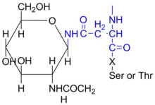
Oligosaccharides that participate in O-linked glycosylation are attached to threonine or serine on the hydroxyl group of the side chain.[7] O-linked glycosylation occurs in the Golgi apparatus, where monosaccharide units are added to a complete polypeptide chain. Cell surface proteins and extracellular proteins are O-glycosylated.[10] Glycosylation sites in O-linked oligosaccharides are determined by the secondary and tertiary structures of the polypeptide, which dictate where glycosyltransferases will add sugars.
Glycosylated biomolecules
Glycoproteins and glycolipids are by definition covalently bonded to carbohydrates. They are very abundant on the surface of the cell, and their interactions contribute to the overall stability of the cell.
Glycoproteins
Glycoproteins have distinct Oligosaccharide structures which have significant effects on many of their properties,[11] affecting critical functions such as antigenicity, solubility, and resistance to proteases. Glycoproteins are relevant as cell-surface receptors, cell-adhesion molecules, immunoglobulins, and tumor antigens.[12]
Glycolipids
Glycolipids are important for cell recognition, and are important for modulating the function of membrane proteins that act as receptors.[13] Glycolipids are lipid molecules bound to oligosaccharides, generally present in the lipid bilayer. Additionally, they can serve as receptors for cellular recognition and cell signaling.[13] The head of the oligosaccharide serves as a binding partner in receptor activity. The binding mechanisms of receptors to the oligosaccharides depends on the composition of the oligosaccharides that are exposed or presented above the surface of the membrane. There is great diversity in the binding mechanisms of glycolipids, which is what makes them such an important target for pathogens as a site for interaction and entrance.[14] For example, the chaperone activity of glycolipids has been studied for its relevance to HIV infection.
Functions
Cell recognition
All cells are coated in either glycoproteins or glycolipids, both of which help determine cell types.[7] Lectins, or proteins that bind carbohydrates, can recognize specific oligosaccharides and provide useful information for cell recognition based on oligosaccharide binding.
An important example of oligosaccharide cell recognition is the role of glycolipids in determining blood types. The various blood types are distinguished by the glycan modification present on the surface of blood cells.[15] These can be visualized using mass spectrometry. The oligosaccharides found on the A, B, and H antigen occur on the non-reducing ends of the oligosaccharide. The H antigen (which indicates an O blood type) serves as a precursor for the A and B antigen.[7] Therefore, a person with A blood type will have the A antigen and H antigen present on the glycolipids of the red blood cell plasma membrane. A person with B blood type will have the B and H antigen present. A person with AB blood type will have A, B, and H antigens present. And finally, a person with O blood type will only have the H antigen present. This means all blood types have the H antigen, which explains why the O blood type is known as the "universal donor".
How do transport vesicles know the final destination of the protein that they are transporting?
Vesicles are directed by many ways, but the two main ways are:
1- The sorting signals encoded in the amino acid sequence of the proteins.
2- The Oligosaccharide attached to the protein.
The sorting signals are recognised by specific receptors that reside in the membranes or surface coasts of budding vesicles, ensuring that the protein is transported to the appropriate destination.
Cell adhesion
Many cells produce specific carbohydrate-binding proteins known as lectins, which mediate cell adhesion with oligosaccharides.[16] Selectins, a family of lectins, mediate certain cell–cell adhesion processes, including those of leukocytes to endothelial cells.[7] In an immune response, endothelial cells can express certain selectins transiently in response to damage or injury to the cells. In response, a reciprocal selectin–oligosaccharide interaction will occur between the two molecules which allows the white blood cell to help eliminate the infection or damage. Protein-Carbohydrate bonding is often mediated by hydrogen bonding and van der Waals forces.
Dietary oligosaccharides
Fructo-oligosaccharides (FOS), which are found in many vegetables, are short chains of fructose molecules. They differ from fructans and inulin, which have a much higher degree of polymerization than FOS and is therefore a polysaccharide, but like fructans and inulin, they are considered soluble dietary fibre. Galactooligosaccharides (GOS), which also occur naturally, consist of short chains of galactose molecules. These compounds cannot be digested in the human small intestine, and instead pass through to the large intestine, where they promote the growth of Bifidobacteria, which are beneficial to gut health.[17]
Mannan oligosaccharides (MOS) are widely used in animal feed to improve gastrointestinal health. They are normally obtained from the yeast cell walls of Saccharomyces cerevisiae. Mannan oligosaccharides differ from other oligosaccharides in that they are not fermentable and their primary mode of actions include agglutination of type-1 fimbria pathogens and immunomodulation[18]
Sources
Oligosaccharides are a component of fibre from plant tissue. FOS and inulin are present in Jerusalem artichoke, burdock, chicory, leeks, onions, and asparagus. Inulin is a significant part of the daily diet of most of the world’s population. FOS can also be synthesized by enzymes of the fungus Aspergillus niger acting on sucrose. GOS is naturally found in soybeans and can be synthesized from lactose. FOS, GOS, and inulin are also sold as nutritional supplements.
See also
- Oligosaccharide synthesis
- Oligosaccharide nomenclature
- Isomaltooligosaccharide (IMO)
References
- "Definition of OLIGOSACCHARIDE". www.merriam-webster.com. Retrieved 2018-10-15.
- Oligosaccharides at the US National Library of Medicine Medical Subject Headings (MeSH)
- Walstra P, Wouters JT, Geurts TJ (2008). Dairy Science and Technology (second ed.). CRC, Taylor & Francis.
- Whitney E, Rolfes SR (2008). Understanding Nutrition (Eleventh ed.). Thomson Wadsworth..
- "Oligosaccharide". Encyclopædia Britannica.
- "Molecular Biology of the Cell. 4th edition". Retrieved 16 August 2018.
- Voet D, Voet J, Pratt C (2013). Fundamentals of Biochemistry: Life at the Molecular Level (4th ed.). Hoboken, NJ: John Wiley & Sons, Inc. ISBN 978-0470-54784-7..
- Varki A (ed.). Essentials of Glycobiology (2nd ed.). Cold Spring Harbor Laboratories Press. ISBN 978-0-87969-770-9..
- Schwarz F, Aebi M (October 2011). "Mechanisms and principles of N-linked protein glycosylation". Current Opinion in Structural Biology. 21 (5): 576–82. doi:10.1016/j.sbi.2011.08.005. PMID 21978957.
- Peter-Katalinić J (2005). "Methods in enzymology: O-glycosylation of proteins". Methods in Enzymology. 405: 139–71. doi:10.1016/S0076-6879(05)05007-X. ISBN 978-0-12-182810-3. PMID 16413314.
- Goochee CF (1992). "Bioprocess factors affecting glycoprotein oligosaccharide structure". Developments in Biological Standardization. 76: 95–104. PMID 1478360.
- Elbein AD (October 1991). "The role of N-linked oligosaccharides in glycoprotein function". Trends in Biotechnology. 9 (10): 346–52. doi:10.1016/0167-7799(91)90117-Z. PMID 1367760.
- Manna M, Róg T, Vattulainen I (August 2014). "The challenges of understanding glycolipid functions: An open outlook based on molecular simulations". Biochimica et Biophysica Acta. 1841 (8): 1130–45. doi:10.1016/j.bbalip.2013.12.016. PMID 24406903.
- Fantini J (2007). "Interaction of proteins with lipid rafts through glycolipid-binding domains: biochemical background and potential therapeutic applications". Current Medicinal Chemistry. 14 (27): 2911–7. doi:10.2174/092986707782360033. PMID 18045136.
- Kailemia MJ, Ruhaak LR, Lebrilla CB, Amster IJ (January 2014). "Oligosaccharide analysis by mass spectrometry: a review of recent developments". Analytical Chemistry. 86 (1): 196–212. doi:10.1021/ac403969n. PMC 3924431. PMID 24313268.
- Feizi T (1993). "Oligosaccharides that mediate mammalian cell–cell adhesion". Current Opinion in Structural Biology. 3 (5): 701–10. doi:10.1016/0959-440X(93)90053-N.
- Moise AM (2017-10-31). The Gut Microbiome: Exploring the Connection between Microbes, Diet, and Health. ABC-CLIO. p. 58. ISBN 978-1-4408-4265-8.
- Smiricky-Tjardes MR, Flickinger EA, Grieshop CM, Bauer LL, Murphy MR, Fahey GC (October 2003). "In vitro fermentation characteristics of selected oligosaccharides by swine fecal microflora". Journal of Animal Science. 81 (10): 2505–14. doi:10.2527/2003.81102505x. PMID 14552378.
External links

Types of carbohydrates | |||||||||||||||
|---|---|---|---|---|---|---|---|---|---|---|---|---|---|---|---|
| General |
| ||||||||||||||
| Geometry |
| ||||||||||||||
| Monosaccharides |
| ||||||||||||||
| Multiple |
| ||||||||||||||
| |||||||||||||||