Cell signaling
In biology, cell signaling (cell signalling in British English) is part of any communication process that governs basic activities of cells and coordinates multiple-cell actions. The ability of cells to perceive and correctly respond to their microenvironment is the basis of development, tissue repair, and immunity, as well as normal tissue homeostasis. Errors in signaling interactions and cellular information processing may cause diseases such as cancer, autoimmunity, and diabetes.[1][2][3] By understanding cell signaling, clinicians may treat diseases more effectively and, theoretically, researchers may develop artificial tissues.[4]
Systems biology studies the underlying structure of cell-signaling networks and how changes in these networks may affect the transmission and flow of information (signal transduction). Such networks are complex systems in their organization and may exhibit a number of emergent properties, including bistability and ultrasensitivity. Analysis of cell-signaling networks requires a combination of experimental and theoretical approaches, including the development and analysis of simulations and modeling.[5][6] Long-range allostery is often a significant component of cell-signaling events.[7]
All cells receive and respond to signals from their surroundings. This is accomplished by a variety of signal molecules that are secreted or expressed on the surface of one cell and bind to a receptor expressed by the other cells, thereby integrating and coordinating the function of the many individual cells that make up organisms. Each cell is programmed to respond to specific extracellular signal molecules. Extracellular signaling usually entails the following steps:
- Synthesis and release of the signaling molecule by the signaling cell;
- Transport of the signal to the target cell;
- Binding of the signal by a specific receptor leading to its activation;
- Initiation of signal-transduction pathways.[8]
Between organisms
Cell signaling has been most extensively studied in the context of human diseases and signaling between cells of a single organism. However, cell signaling may also occur between the cells of two different organisms. In many mammals, early embryo cells exchange signals with cells of the uterus.[9] In the human gastrointestinal tract, bacteria exchange signals with each other and with human epithelial and immune system cells.[10] For the yeast Saccharomyces cerevisiae during mating, some cells send a peptide signal (mating factor pheromones) into their environment. The mating factor peptide may bind to a cell surface receptor on other yeast cells and induce them to prepare for mating.[11]
Classification
Cell signaling can be classified as either mechanical or biochemical based on the type of the signal. Mechanical signals are the forces exerted on the cell and the forces produced by the cell. These forces can both be sensed and responded to by the cells.[12] Biochemical signals are biochemical molecules such as proteins, lipids, ions, and gases. These signals can be categorized based on the distance between signaling and responder cells. Signaling within, between, and amongst cells is subdivided into the following classifications:
- Intracrine signals are produced by the target cell that stay within the target cell.
- Autocrine signals are produced by the target cell, are secreted, and affect the target cell itself via receptors. Sometimes autocrine cells can target cells close by if they are the same type of cell as the emitting cell. An example of this are immune cells.
- Juxtacrine signals target adjacent (touching) cells. These signals are transmitted along cell membranes via protein or lipid components integral to the membrane and are capable of affecting either the emitting cell or cells immediately adjacent.
- Paracrine signals target cells in the vicinity of the emitting cell. Neurotransmitters represent an example.
- Endocrine signals target distant cells. Endocrine cells produce hormones that travel through the blood to reach all parts of the body.
Cells communicate with each other via direct contact (juxtacrine signaling), over short distances (paracrine signaling), or over large distances and/or scales (endocrine signaling).
Some cell–cell communication requires direct cell–cell contact. Some cells can form gap junctions that connect their cytoplasm to the cytoplasm of adjacent cells. In cardiac muscle, gap junctions between adjacent cells allow for action potential propagation from the cardiac pacemaker region of the heart to spread and coordinate the contraction of the heart.
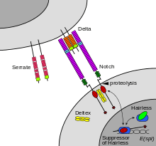
The notch signaling mechanism is an example of juxtacrine signaling (also known as contact-dependent signaling) in which two adjacent cells must make physical contact in order to communicate. This requirement for direct contact allows for very precise control of cell differentiation during embryonic development. In the worm Caenorhabditis elegans, two cells of the developing gonad each have an equal chance of terminally differentiating or becoming a uterine precursor cell that continues to divide. The choice of which cell continues to divide is controlled by competition of cell surface signals. One cell will happen to produce more of a cell surface protein that activates the Notch receptor on the adjacent cell. This activates a feedback loop or system that reduces Notch expression in the cell that will differentiate and that increases Notch on the surface of the cell that continues as a stem cell.[13]
Many cell signals are carried by molecules that are released by one cell and move to make contact with another cell. Endocrine signals are called hormones. Hormones are produced by endocrine cells and they travel through the blood to reach all parts of the body. Specificity of signaling can be controlled if only some cells can respond to a particular hormone. Paracrine signals such as retinoic acid target only cells in the vicinity of the emitting cell.[14] Neurotransmitters represent another example of a paracrine signal. Some signaling molecules can function as both a hormone and a neurotransmitter. For example, epinephrine and norepinephrine can function as hormones when released from the adrenal gland and are transported to the heart by way of the blood stream. Norepinephrine can also be produced by neurons to function as a neurotransmitter within the brain.[15] Estrogen can be released by the ovary and function as a hormone or act locally via paracrine or autocrine signaling.[16] Active species of oxygen and nitric oxide can also act as cellular messengers. This process is dubbed redox signaling.
In multicellular organisms
In a multicellular organism, signaling between cells occurs either through release into the extracellular space, divided in paracrine signaling (over short distances) and endocrine signaling (over long distances), or by direct contact, known as juxtacrine signaling.[17] Autocrine signaling is a special case of paracrine signaling where the secreting cell has the ability to respond to the secreted signaling molecule.[18] Synaptic signaling is a special case of paracrine signaling (for chemical synapses) or juxtacrine signaling (for electrical synapses) between neurons and target cells. Signaling molecules interact with a target cell as a ligand to cell surface receptors, and/or by entering into the cell through its membrane or endocytosis for intracrine signaling. This generally results in the activation of second messengers, leading to various physiological effects.
A particular molecule is generally used in diverse modes of signaling, and therefore a classification by mode of signaling is not possible. At least three important classes of signaling molecules are widely recognized, although non-exhaustive and with imprecise boundaries, as such membership is non-exclusive and depends on the context:
- Hormones are the major signaling molecules of the endocrine system, though they often regulate each other's secretion via local signaling (e.g. islet of Langerhans cells), and most are also expressed in tissues for local purposes (e.g. angiotensin) or failing that, structurally related molecules are (e.g. PTHrP).
- Neurotransmitters are signaling molecules of the nervous system, also including neuropeptides and neuromodulators. Neurotransmitters like the catecholamines are also secreted by the endocrine system into the systemic circulation.
- Cytokines are signaling molecules of the immune system, with a primary paracrine or juxtacrine role, though they can during significant immune responses have a strong presence in the circulation, with systemic effect (altering iron metabolism or body temperature). Growth factors can be considered as cytokines or a different class.
Signaling molecules can belong to several chemical classes: lipids, phospholipids, amino acids, monoamines, proteins, glycoproteins, or gases. Signaling molecules binding surface receptors are generally large and hydrophilic (e.g. TRH, Vasopressin, Acetylcholine), while those entering the cell are generally small and hydrophobic (e.g. glucocorticoids, thyroid hormones, cholecalciferol, retinoic acid), but important exceptions to both are numerous, and a same molecule can act both via surface receptor or in an intracrine manner to different effects.[18] In intracrine signaling, once inside the cell, a signaling molecule can bind to intracellular receptors, other elements, or stimulate enzyme activity (e.g. gasses). The intracrine action of peptide hormones remains a subject of debate.[19]
Hydrogen sulfide is produced in small amounts by some cells of the human body and has a number of biological signaling functions. Only two other such gases are currently known to act as signaling molecules in the human body: nitric oxide and carbon monoxide.[20]
Signaling receptors
Cells receive information from their neighbors through a class of proteins known as receptors. Notch is a cell surface protein that functions as a receptor. Animals have a small set of genes that code for signaling proteins that interact specifically with Notch receptors and stimulate a response in cells that express Notch on their surface. Molecules that activate (or, in some cases, inhibit) receptors can be classified as hormones, neurotransmitters, cytokines, and growth factors, in general called receptor ligands. Ligand receptor interactions such as that of the Notch receptor interaction, are known to be the main interactions responsible for cell signaling mechanisms and communication.[21]
As shown in Figure 2 (above; left), notch acts as a receptor for ligands that are expressed on adjacent cells. While some receptors are cell-surface proteins, others are found inside cells. For example, estrogen is a hydrophobic molecule that can pass through the lipid bilayer of the membranes. As part of the endocrine system, intracellular estrogen receptors from a variety of cell types can be activated by estrogen produced in the ovaries.
A number of transmembrane receptors[22][23] for small molecules and peptide hormones,[24] as well as intracellular receptors for steroid hormones exist, giving cells the ability to respond to a great number of hormonal and pharmacological stimuli. In diseases, often, proteins that interact with receptors are aberrantly activated, resulting in constitutively activated downstream signals.[25]
For several types of intercellular signaling molecules that are unable to permeate the hydrophobic cell membrane due to their hydrophilic nature, the target receptor is expressed on the membrane. When such a signaling molecule activates its receptor, the signal is carried into the cell usually by means of a second messenger such as cAMP.[26][27]
Signaling pathways
In some cases, receptor activation caused by ligand binding to a receptor is directly coupled to the cell's response to the ligand. For example, the neurotransmitter GABA can activate a cell surface receptor that is part of an ion channel. GABA binding to a GABAA receptor on a neuron opens a chloride-selective ion channel that is part of the receptor. GABAA receptor activation allows negatively charged chloride ions to move into the neuron, which inhibits the ability of the neuron to produce action potentials. However, for many cell surface receptors, ligand-receptor interactions are not directly linked to the cell's response. The activated receptor must first interact with other proteins inside the cell before the ultimate physiological effect of the ligand on the cell's behavior is produced. Often, the behavior of a chain of several interacting cell proteins is altered following receptor activation. The entire set of cell changes induced by receptor activation is called a signal transduction mechanism or pathway.[28]
In the case of Notch-mediated signaling, the signal transduction mechanism can be relatively simple. As shown in Figure 2, the activation of Notch can cause the Notch protein to be altered by a protease. Part of the Notch protein is released from the cell surface membrane and takes part in gene regulation. Cell signaling research involves studying the spatial and temporal dynamics of both receptors and the components of signaling pathways that are activated by receptors in various cell types.[29][30] Emerging methods for single-cell mass-spectrometry analysis promise to enable studying signal transduction with single-cell resolution.[31]
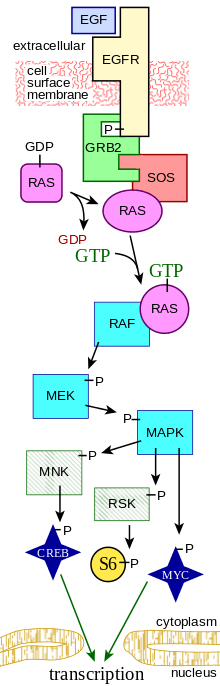
A more complex signal transduction pathway is shown in Figure 3. This pathway involves changes of protein–protein interactions inside the cell, induced by an external signal. Many growth factors bind to receptors at the cell surface and stimulate cells to progress through the cell cycle and divide. Several of these receptors are kinases that start to phosphorylate themselves and other proteins when binding to a ligand. This phosphorylation can generate a binding site for a different protein and thus induce protein–protein interaction. In Figure 3, the ligand (called epidermal growth factor, or EGF) binds to the receptor (called EGFR). This activates the receptor to phosphorylate itself. The phosphorylated receptor binds to an adaptor protein (GRB2), which couples the signal to further downstream signaling processes. For example, one of the signal transduction pathways that are activated is called the mitogen-activated protein kinase (MAPK) pathway. The signal transduction component labeled as "MAPK" in the pathway was originally called "ERK," so the pathway is called the MAPK/ERK pathway. The MAPK protein is an enzyme, a protein kinase that can attach phosphate to target proteins such as the transcription factor MYC and, thus, alter gene transcription and, ultimately, cell cycle progression. Many cellular proteins are activated downstream of the growth factor receptors (such as EGFR) that initiate this signal transduction pathway.
Some signaling transduction pathways respond differently, depending on the amount of signaling received by the cell. For instance, the hedgehog protein activates different genes, depending on the amount of hedgehog protein present.
Complex multi-component signal transduction pathways provide opportunities for feedback, signal amplification, and interactions inside one cell between multiple signals and signaling pathways.
Intra- and inter-species signaling
Molecular signaling can occur between different organisms, whether unicellular or multicellular. The emitting organism produces the signaling molecule, secretes it into the environment, where it diffuses, and it is sensed or internalized by the receiving organism. In some cases of interspecies signaling, the emitting organism can actually be a host of the receiving organism, or vice versa.
Intraspecies signaling occurs especially in bacteria, yeast, social insects, but also many vertebrates. The signaling molecules used by multicellular organisms are often called pheromones. They can have such purposes as alerting against danger, indicating food supply, or assisting in reproduction.[32] In unicellular organisms such as bacteria, signaling can be used to 'activate' peers from a dormant state, enhance virulence, defend against bacteriophages, etc.[33] In quorum sensing, which is also found in social insects, the multiplicity of individual signals has the potentiality to create a positive feedback loop, generating coordinated response. In this context, the signaling molecules are called autoinducers.[34][35][36] This signaling mechanism may have been involved in evolution from unicellular to multicellular organisms.[34][37] Bacteria also use contact-dependent signaling, notably to limit their growth.[38]
Molecular signaling can also occur between individuals of different species. This has been particularly studied in bacteria.[39][40][41] Different bacterial species can coordinate to colonize a host and participate in common quorum sensing.[42] Therapeutic strategies to disrupt this phenomenon are being investigated.[43][44] Interactions mediated through signaling molecules are also thought to occur between the gut flora and their host, as part of their commensal or symbiotic relationship.[44][45] Gram negative microbes deploy bacterial outer membrane vesicles for intra- and inter-species signaling in natural environments and at the host-pathogen interface.
Additionally, interspecies signaling occurs between multicellular organisms. In Vespa mandarinia, individuals release a scent that directs the colony to a food source.[46]
Computational models
Recent approaches to better understand elements of pathway crosstalk, complex ligand-receptor binding, and signaling network dynamics have been aided by the use of systems biology approaches.[47] Computational models often take aim at compiling information from published literature to generate a coherent set of signaling components and their associated interactions.[48] The development of computational models allows for a more in-depth probing of cell signaling pathways at a global level by manipulating different variables and systemically evaluating the resulting response.[49] The use of analytical models for the study of signal transduction has been heavily applied in the fields of pharmacology and drug discovery to assess receptor-ligand interactions and pharmacokinetics as well as the flow of metabolites in large networks.[5] A commonly applied strategy to model cell signaling mechanisms is through the use of ordinary differential equation (ODE) models by expressing the time-dependent concentration of a signaling molecule as a function of other molecules downstream and/or upstream within the pathway.[50] ODE models have already been applied for dynamic analysis of the Mitogen-activated protein kinase, Estrogen receptor alpha, and MTOR signaling pathways among numerous others.[51][52][53]
See also
- Biosemiotics
- Molecular cellular cognition
- Cellular communication (biology)
- Crosstalk (biology)
- Bacterial outer membrane vesicles
- Membrane vesicle trafficking
- Host-pathogen interface
- MAPK signaling pathway
- Wnt signaling pathway
- Hedgehog signaling pathway
- Retinoic acid
- TGF beta signaling pathway
- JAK-STAT signaling pathway
- cAMP-dependent pathway
- Imd pathway
- Protein dynamics
- Signal transduction
- Systems biology
- Lipid signaling
- Redox signaling
- Cell Signaling Technology, an antibody development and production company
- Netpath – A curated resource of signal transduction pathways in humans
- Nanoscale networking – leveraging biological signaling to construct ad hoc in vivo communication networks
- Soliton model in neuroscience—Physical communication via sound waves in membranes
References
- Vlahopoulos SA, Cen O, Hengen N, Agan J, Moschovi M, Critselis E, Adamaki M, Bacopoulou F, Copland JA, Boldogh I, Karin M, Chrousos GP (August 2015). "Dynamic aberrant NF-κB spurs tumorigenesis: a new model encompassing the microenvironment". Cytokine & Growth Factor Reviews. 26 (4): 389–403. doi:10.1016/j.cytogfr.2015.06.001. PMC 4526340. PMID 26119834.
- Wang K, Grivennikov SI, Karin M (April 2013). "Implications of anti-cytokine therapy in colorectal cancer and autoimmune diseases". Annals of the Rheumatic Diseases. 72 Suppl 2: ii100–3. doi:10.1136/annrheumdis-2012-202201. PMID 23253923.
We have shown interleukin (IL)-6 to be an important tumour promoter in early colitis-associated cancer (CAC).
- Solinas G, Vilcu C, Neels JG, Bandyopadhyay GK, Luo JL, Naugler W, Grivennikov S, Wynshaw-Boris A, Scadeng M, Olefsky JM, Karin M (November 2007). "JNK1 in hematopoietically derived cells contributes to diet-induced inflammation and insulin resistance without affecting obesity". Cell Metabolism. 6 (5): 386–97. doi:10.1016/j.cmet.2007.09.011. PMID 17983584.
Activation of JNKs (mainly JNK1) in insulin target cells results in phosphorylation of insulin receptor substrates (IRSs) at serine and threonine residues that inhibit insulin signaling.
- Smith RJ, Koobatian MT, Shahini A, Swartz DD, Andreadis ST (May 2015). "Capture of endothelial cells under flow using immobilized vascular endothelial growth factor". Biomaterials. 51: 303–312. doi:10.1016/j.biomaterials.2015.02.025. PMC 4361797. PMID 25771020.
- Eungdamrong NJ, Iyengar R (June 2004). "Modeling cell signaling networks". Biology of the Cell. 96 (5): 355–62. doi:10.1016/j.biolcel.2004.03.004. PMC 3620715. PMID 15207904.
- Dong P, Maddali MV, Srimani JK, Thélot F, Nevins JR, Mathey-Prevot B, You L (September 2014). "Division of labour between Myc and G1 cyclins in cell cycle commitment and pace control". Nature Communications. 5: 4750. Bibcode:2014NatCo...5E4750D. doi:10.1038/ncomms5750. PMC 4164785. PMID 25175461.
- Bu Z, Callaway DJ (2011). "Proteins move! Protein dynamics and long-range allostery in cell signaling". Protein Structure and Diseases. Advances in Protein Chemistry and Structural Biology. 83. pp. 163–221. doi:10.1016/B978-0-12-381262-9.00005-7. ISBN 978-0-12-381262-9. PMID 21570668.
- Kumar P, Mina U (2014). Life science fundamental and practice part I. New Delhi, India: Pathfinder Publication.
- Mohamed OA, Jonnaert M, Labelle-Dumais C, Kuroda K, Clarke HJ, Dufort D (June 2005). "Uterine Wnt/beta-catenin signaling is required for implantation". Proceedings of the National Academy of Sciences of the United States of America. 102 (24): 8579–84. Bibcode:2005PNAS..102.8579M. doi:10.1073/pnas.0500612102. PMC 1150820. PMID 15930138.
- Clarke MB, Sperandio V (June 2005). "Events at the host-microbial interface of the gastrointestinal tract III. Cell-to-cell signaling among microbial flora, host, and pathogens: there is a whole lot of talking going on". American Journal of Physiology. Gastrointestinal and Liver Physiology. 288 (6): G1105–9. doi:10.1152/ajpgi.00572.2004. PMID 15890712.
- Lin JC, Duell K, Konopka JB (March 2004). "A microdomain formed by the extracellular ends of the transmembrane domains promotes activation of the G protein-coupled alpha-factor receptor". Molecular and Cellular Biology. 24 (5): 2041–51. doi:10.1128/MCB.24.5.2041-2051.2004. PMC 350546. PMID 14966283.
- Miller CJ, Davidson LA (October 2013). "The interplay between cell signalling and mechanics in developmental processes". Nature Reviews Genetics. 14 (10): 733–44. doi:10.1038/nrg3513. PMC 4056017. PMID 24045690.
- Greenwald I (June 1998). "LIN-12/Notch signaling: lessons from worms and flies". Genes & Development. 12 (12): 1751–62. doi:10.1101/gad.12.12.1751. PMID 9637676.
- Duester G (September 2008). "Retinoic acid synthesis and signaling during early organogenesis". Cell. 134 (6): 921–31. doi:10.1016/j.cell.2008.09.002. PMC 2632951. PMID 18805086.
- Cartford MC, Samec A, Fister M, Bickford PC (2004). "Cerebellar norepinephrine modulates learning of delay classical eyeblink conditioning: evidence for post-synaptic signaling via PKA". Learning & Memory. 11 (6): 732–7. doi:10.1101/lm.83104. PMC 534701. PMID 15537737.
- Jesmin S, Mowa CN, Sakuma I, Matsuda N, Togashi H, Yoshioka M, Hattori Y, Kitabatake A (October 2004). "Aromatase is abundantly expressed by neonatal rat penis but downregulated in adulthood". Journal of Molecular Endocrinology. 33 (2): 343–59. doi:10.1677/jme.1.01548. PMID 15525594.
- Gilbert SF (2000). "Juxtacrine Signaling". In NCBI bookshelf (ed.). Developmental biology (6. ed.). Sunderland, Mass.: Sinauer Assoc. ISBN 978-0878932436.
- Alberts B, Johnson A, Lewis J, et al. (2002). "General Principles of Cell Communication". In NCBI bookshelf (ed.). Molecular biology of the cell (4th ed.). New York: Garland Science. ISBN 978-0815332183.
- Re R (October 1999). "The nature of intracrine peptide hormone action". Hypertension. 34 (4 Pt 1): 534–8. CiteSeerX 10.1.1.326.4497. doi:10.1161/01.HYP.34.4.534. PMID 10523322.
- Cooper GM, Hausman RE (2000). "Signaling Molecules and Their Receptors". In NCBI bookshelf (ed.). The cell : a molecular approach (2nd ed.). Washington, D.C.: ASM Press. ISBN 978-0878933006.
- Cooper GM (2000). "Functions of Cell Surface Receptors.". The Cell: A Molecular Approach (2nd ed.). Sunderland (MA): Sinauer Associates.
- Domazet I, Holleran BJ, Martin SS, Lavigne P, Leduc R, Escher E, Guillemette G (May 2009). "The second transmembrane domain of the human type 1 angiotensin II receptor participates in the formation of the ligand binding pocket and undergoes integral pivoting movement during the process of receptor activation". The Journal of Biological Chemistry. 284 (18): 11922–9. doi:10.1074/jbc.M808113200. PMC 2673261. PMID 19276075.
- Hislop JN, Henry AG, Marchese A, von Zastrow M (July 2009). "Ubiquitination regulates proteolytic processing of G protein-coupled receptors after their sorting to lysosomes". The Journal of Biological Chemistry. 284 (29): 19361–70. doi:10.1074/jbc.M109.001644. PMC 2740561. PMID 19433584.
- Meng H, Zhang X, Hankenson KD, Wang MM (March 2009). "Thrombospondin 2 potentiates notch3/jagged1 signaling". The Journal of Biological Chemistry. 284 (12): 7866–74. doi:10.1074/jbc.M803650200. PMC 2658079. PMID 19147503.
- Copland JA, Sheffield-Moore M, Koldzic-Zivanovic N, Gentry S, Lamprou G, Tzortzatou-Stathopoulou F, Zoumpourlis V, Urban RJ, Vlahopoulos SA (June 2009). "Sex steroid receptors in skeletal differentiation and epithelial neoplasia: is tissue-specific intervention possible?". BioEssays. 31 (6): 629–41. doi:10.1002/bies.200800138. PMID 19382224.
- Goh SL, Looi Y, Shen H, Fang J, Bodner C, Houle M, Ng AC, Screaton RA, Featherstone M (July 2009). "Transcriptional activation by MEIS1A in response to protein kinase A signaling requires the transducers of regulated CREB family of CREB co-activators". The Journal of Biological Chemistry. 284 (28): 18904–12. doi:10.1074/jbc.M109.005090. PMC 2707216. PMID 19473990.
- Wojtal KA, Hoekstra D, van Ijzendoorn SC (February 2008). "cAMP-dependent protein kinase A and the dynamics of epithelial cell surface domains: moving membranes to keep in shape" (PDF). BioEssays. 30 (2): 146–55. doi:10.1002/bies.20705. PMID 18200529.
- Dinasarapu AR, Saunders B, Ozerlat I, Azam K, Subramaniam S (June 2011). "Signaling gateway molecule pages--a data model perspective". Bioinformatics. 27 (12): 1736–8. doi:10.1093/bioinformatics/btr190. PMC 3106186. PMID 21505029.
- Ferrell JE, Machleder EM (May 1998). "The biochemical basis of an all-or-none cell fate switch in Xenopus oocytes". Science. 280 (5365): 895–8. doi:10.1126/science.280.5365.895. PMID 9572732.
- Slavov N, Carey J, Linse S (April 2013). "Calmodulin transduces Ca2+ oscillations into differential regulation of its target proteins". ACS Chemical Neuroscience. 4 (4): 601–12. doi:10.1021/cn300218d. PMC 3629746. PMID 23384199.
- Slavov N (January 2020). "Unpicking the proteome in single cells". Science. 367 (6477): 512–513. doi:10.1126/science.aaz6695. PMC 7029782. PMID 32001644.
- Tirindelli R, Dibattista M, Pifferi S, Menini A (July 2009). "From pheromones to behavior". Physiological Reviews. 89 (3): 921–56. CiteSeerX 10.1.1.460.5566. doi:10.1152/physrev.00037.2008. PMID 19584317.
- Mukamolova GV, Kaprelyants AS, Young DI, Young M, Kell DB (July 1998). "A bacterial cytokine". Proceedings of the National Academy of Sciences of the United States of America. 95 (15): 8916–21. Bibcode:1998PNAS...95.8916M. doi:10.1073/pnas.95.15.8916. PMC 21177. PMID 9671779.
- Miller MB, Bassler BL (1 October 2001). "Quorum sensing in bacteria". Annual Review of Microbiology. 55 (1): 165–99. doi:10.1146/annurev.micro.55.1.165. PMID 11544353.
- Kaper JB, Sperandio V (June 2005). "Bacterial cell-to-cell signaling in the gastrointestinal tract". Infection and Immunity. 73 (6): 3197–209. doi:10.1128/IAI.73.6.3197-3209.2005. PMC 1111840. PMID 15908344.
- Camilli A, Bassler BL (February 2006). "Bacterial small-molecule signaling pathways". Science. 311 (5764): 1113–6. Bibcode:2006Sci...311.1113C. doi:10.1126/science.1121357. PMC 2776824. PMID 16497924.
- Stoka AM (June 1999). "Phylogeny and evolution of chemical communication: an endocrine approach". Journal of Molecular Endocrinology. 22 (3): 207–25. doi:10.1677/jme.0.0220207. PMID 10343281.
- Blango MG, Mulvey MA (April 2009). "Bacterial landlines: contact-dependent signaling in bacterial populations". Current Opinion in Microbiology. 12 (2): 177–81. doi:10.1016/j.mib.2009.01.011. PMC 2668724. PMID 19246237.
- Shank EA, Kolter R (April 2009). "New developments in microbial interspecies signaling". Current Opinion in Microbiology. 12 (2): 205–14. doi:10.1016/j.mib.2009.01.003. PMC 2709175. PMID 19251475.
- Ryan RP, Dow JM (July 2008). "Diffusible signals and interspecies communication in bacteria". Microbiology. 154 (Pt 7): 1845–58. doi:10.1099/mic.0.2008/017871-0. PMID 18599814.
- Ryan RP, Dow JM (March 2011). "Communication with a growing family: diffusible signal factor (DSF) signaling in bacteria". Trends in Microbiology. 19 (3): 145–52. doi:10.1016/j.tim.2010.12.003. PMID 21227698.
- Déziel E, Lépine F, Milot S, He J, Mindrinos MN, Tompkins RG, Rahme LG (February 2004). "Analysis of Pseudomonas aeruginosa 4-hydroxy-2-alkylquinolines (HAQs) reveals a role for 4-hydroxy-2-heptylquinoline in cell-to-cell communication". Proceedings of the National Academy of Sciences of the United States of America. 101 (5): 1339–44. Bibcode:2004PNAS..101.1339D. doi:10.1073/pnas.0307694100. PMC 337054. PMID 14739337.
- Federle MJ, Bassler BL (November 2003). "Interspecies communication in bacteria". The Journal of Clinical Investigation. 112 (9): 1291–9. doi:10.1172/JCI20195. PMC 228483. PMID 14597753.
- Sperandio V, Torres AG, Jarvis B, Nataro JP, Kaper JB (July 2003). "Bacteria-host communication: the language of hormones". Proceedings of the National Academy of Sciences of the United States of America. 100 (15): 8951–6. Bibcode:2003PNAS..100.8951S. doi:10.1073/pnas.1537100100. PMC 166419. PMID 12847292.
- Hooper LV, Gordon JI (May 2001). "Commensal host-bacterial relationships in the gut". Science. 292 (5519): 1115–8. Bibcode:2001Sci...292.1115H. doi:10.1126/science.1058709. PMID 11352068.
- Schueller TI, Nordheim EV, Taylor BJ, Jeanne RL (November 2010). "The cues have it; nest-based, cue-mediated recruitment to carbohydrate resources in a swarm-founding social wasp". Die Naturwissenschaften. 97 (11): 1017–22. Bibcode:2010NW.....97.1017S. doi:10.1007/s00114-010-0712-9. PMID 20821186.
- Chen RE, Thorner J (2005). "Systems biology approaches in cell signaling research". Genome Biology. 6 (10): 235. doi:10.1186/gb-2005-6-10-235. PMC 1257459. PMID 16207364.
- Hughey JJ, Lee TK, Covert MW (2010). "Computational modeling of mammalian signaling networks". Wiley Interdisciplinary Reviews: Systems Biology and Medicine. 2 (2): 194–209. doi:10.1002/wsbm.52. PMC 3105527. PMID 20836022.
- Rangamani P, Iyengar R (2008). "Modelling cellular signalling systems". Essays in Biochemistry. 45: 83–94. doi:10.1042/BSE0450083. PMC 3270941. PMID 18793125.
- Poupon A, Reiter E (January 2014). Computational Models to Decipher Cell-Signaling Pathways. Cellular Endocrinology in Health and Disease 2014. pp. 269–284. doi:10.1016/B978-0-12-408134-5.00017-2. ISBN 9780124081345.
- Kolch W, Calder M, Gilbert D (March 2005). "When kinases meet mathematics: the systems biology of MAPK signalling". FEBS Letters. 579 (8): 1891–5. CiteSeerX 10.1.1.584.6262. doi:10.1016/j.febslet.2005.02.002. PMID 15763569.
- Tian D, Solodin NM, Rajbhandari P, Bjorklund K, Alarid ET, Kreeger PK (May 2015). "A kinetic model identifies phosphorylated estrogen receptor-α (ERα) as a critical regulator of ERα dynamics in breast cancer". FASEB Journal. 29 (5): 2022–31. doi:10.1096/fj.14-265637. PMC 4415015. PMID 25648997.
- Sulaimanov N, Klose M, Busch H, Boerries M (July 2017). "Understanding the mTOR signaling pathway via mathematical modeling". Wiley Interdisciplinary Reviews: Systems Biology and Medicine. 9 (4): e1379. doi:10.1002/wsbm.1379. PMC 5573916. PMID 28186392.
External links
- NCI-Nature Pathway Interaction Database: authoritative information about signaling pathways in human cells.
- Intercellular+Signaling+Peptides+and+Proteins at the US National Library of Medicine Medical Subject Headings (MeSH)
- Cell+Communication at the US National Library of Medicine Medical Subject Headings (MeSH)
Metabolism map | ||
|---|---|---|
Single lines: pathways common to most lifeforms. Double lines: pathways not in humans (occurs in e.g. plants, fungi, prokaryotes). | ||
.svg.png)