Cerebral infarction
A cerebral infarction is an area of necrotic tissue in the brain resulting from a blockage or narrowing in the arteries supplying blood and oxygen to the brain. The restricted oxygen due to the restricted blood supply causes an ischemic stroke that can result in an infarction if the blood flow is not restored within a relatively short period of time. The blockage can be due to a thrombus, an embolus or an atheromatous stenosis of one or more arteries.[1] Which arteries are problematic will determine which areas of the brain are affected (infarcted). These varying infarcts will produce different symptoms and outcomes. About one third will prove fatal.
| Cerebral infarct | |
|---|---|
 | |
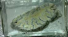
| CT scan slice of the brain showing a right-hemispheric cerebral infarct (left side of image). | |
| Specialty | Neurology |
Classification
There are various classification systems[2] for a cerebral infarction, some of which are described below.
- The Oxford Community Stroke Project classification (OCSP, also known as the Bamford or Oxford classification) relies primarily on the initial symptoms. Based on the extent of the symptoms, the stroke episode is classified as total anterior circulation infarct (TACI), partial anterior circulation infarct (PACI), lacunar infarct (LACI) or posterior circulation infarct (POCI). These four entities predict the extent of the stroke, the area of the brain affected, the underlying cause, and the prognosis.[3][4]
- The TOAST (Trial of Org 10172 in Acute Stroke Treatment) classification is based on clinical symptoms as well as results of further investigations; on this basis, a stroke is classified as being due to (1) thrombosis or embolism due to atherosclerosis of a large artery, (2) embolism of cardiac origin, (3) occlusion of a small blood vessel, (4) other determined cause, (5) undetermined cause (two possible causes, no cause identified, or incomplete investigation).[5]
Symptoms
Symptoms of cerebral infarction are determined by the parts of the brain affected. If the infarct is located in primary motor cortex, contralateral hemiparesis is said to occur. With brainstem localization, brainstem syndromes are typical: Wallenberg's syndrome, Weber's syndrome, Millard–Gubler syndrome, Benedikt syndrome or others.
Infarctions will result in weakness and loss of sensation on the opposite side of the body. Physical examination of the head area will reveal abnormal pupil dilation, light reaction and lack of eye movement on opposite side. If the infarction occurs on the left side brain, speech will be slurred. Reflexes may be aggravated as well.
Risk factors
Major risk factors for cerebral infarction are generally the same as for atherosclerosis: high blood pressure, Diabetes mellitus, tobacco smoking, obesity, and dyslipidemia.[6] The American Heart Association/American Stroke Association (AHA/ASA) recommends controlling these risk factors in order to prevent stroke.[7] The AHA/ASA guidelines also provide information on how to prevent stroke if someone has more specific concerns, such as Sickle-cell disease or pregnancy. It is also possible to calculate the risk of stroke in the next decade based on information gathered through the Framingham Heart Study.[8]
Pathophysiology
Whether a cerebral infarction is thrombotic or embolic based, its pathophysiology, or the observed conditions and underlying mechanisms of the disease. In thrombotic ischemic stroke, a thrombus forms and blocks blood flow.[10] A thrombus forms when the endothelium is activated by a variety of signals to result in platelet aggregation in the artery. This clump of platelets interacts with fibrin to form a platelet plug.[11] This platelet plug grows into a thrombus, resulting in a stenotic artery. Thrombotic ischemia can occur in large or small blood vessels. In large vessels, the most common causes of thrombi are atherosclerosis and vasoconstriction. In small vessels, the most common cause is lipohyalinosis.[12] Lipohyalinosis is when high blood pressure and aging causes a build-up of fatty hyaline matter in blood vessels. Atheroma formation can also cause small vessel thrombotic ischemic stroke.[13] Occlusion of blood flow generates a depolarization of neurons that generate a wave called spreading depolarization,[14] that moves from the affected core to the penumbra and healthy brain producing further depolarization, neuronal dead and neurologic symptoms.[15]
An embolic stroke refers to the blockage of an artery by an embolus, a traveling particle or debris in the arterial bloodstream originating elsewhere. An embolus is most frequently a thrombus, but it can also be a number of other substances including fat (e.g. from bone marrow in a broken bone), air, cancer cells or clumps of bacteria (usually from infectious endocarditis). The embolus may be of cardiac origin due to Atrial fibrillation, Patent foramen ovale or from atherosclerotic plaque of another (or the same) large artery. Cerebral artery gas embolism (e.g. during ascent from a SCUBA dive) is also a possible cause of infarction (Levvett & Millar, 2008)
Diagnosis
Computed tomography (CT) and MRI scanning will show damaged area in the brain, showing that the symptoms were not caused by a tumor, subdural hematoma or other brain disorder. The blockage will also appear on the angiogram.
Treatment
In last decade, similar to myocardial infarction treatment, thrombolytic drugs were introduced in the therapy of cerebral infarction. The use of intravenous rtPA therapy can be advocated in patients who arrive to stroke unit and can be fully evaluated within 3 h of the onset.
If cerebral infarction is caused by a thrombus occluding blood flow to an artery supplying the brain, definitive therapy is aimed at removing the blockage by breaking the clot down (thrombolysis), or by removing it mechanically (thrombectomy). The more rapidly blood flow is restored to the brain, the fewer brain cells die.[16] In increasing numbers of primary stroke centers, pharmacologic thrombolysis with the drug tissue plasminogen activator (tPA), is used to dissolve the clot and unblock the artery. Another intervention for acute cerebral ischaemia is removal of the offending thrombus directly. This is accomplished by inserting a catheter into the femoral artery, directing it into the cerebral circulation, and deploying a corkscrew-like device to ensnare the clot, which is then withdrawn from the body. Mechanical embolectomy devices have been demonstrated effective at restoring blood flow in patients who were unable to receive thrombolytic drugs or for whom the drugs were ineffective,[17][18][19][20] though no differences have been found between newer and older versions of the devices.[21] The devices have only been tested on patients treated with mechanical clot embolectomy within eight hours of the onset of symptoms.
Angioplasty and stenting have begun to be looked at as possible viable options in treatment of acute cerebral ischaemia. In a systematic review of six uncontrolled, single-center trials, involving a total of 300 patients, of intra-cranial stenting in symptomatic intracranial arterial stenosis, the rate of technical success (reduction to stenosis of <50%) ranged from 90-98%, and the rate of major peri-procedural complications ranged from 4-10%. The rates of restenosis and/or stroke following the treatment were also favorable.[22] This data suggests that a large, randomized controlled trial is needed to more completely evaluate the possible therapeutic advantage of this treatment.
If studies show carotid stenosis, and the patient has residual function in the affected side, carotid endarterectomy (surgical removal of the stenosis) may decrease the risk of recurrence if performed rapidly after cerebral infarction. Carotid endarterectomy is also indicated to decrease the risk of cerebral infarction for symptomatic carotid stenosis (>70 to 80% reduction in diameter).[23]
In tissue losses that are not immediately fatal, the best course of action is to make every effort to restore impairments through physical therapy, cognitive therapy, occupational therapy, speech therapy and exercise.
References
- Ropper, Allan H.; Adams, Raymond Delacy; Brown, Robert F.; Victor, Maurice (2005). Adams and Victor's principles of neurology. New York: McGraw-Hill Medical Pub. Division. pp. 686–704. ISBN 0-07-141620-X.
- Amarenco, P.; Bogousslavsky, J.; Caplan, L.R.; Donnan, G.A.; Hennerici, M.G. (2009). "Classification of Stroke Subtypes". Cerebrovascular Diseases. 27 (5): 493–501. doi:10.1159/000210432. ISSN 1015-9770. PMID 19342825.
- Bamford J, Sandercock P, Dennis M, Burn J, Warlow C (June 1991). "Classification and natural history of clinically identifiable subtypes of cerebral infarction". Lancet. 337 (8756): 1521–6. doi:10.1016/0140-6736(91)93206-O. PMID 1675378. Later publications distinguish between "syndrome" and "infarct", based on evidence from imaging. "Syndrome" may be replaced by "hemorrhage" if imaging demonstrates a bleed. See Internet Stroke Center. "Oxford Stroke Scale". Retrieved 2008-11-14.
- Bamford JM (2000). "The role of the clinical examination in the subclassification of stroke". Cerebrovasc. Dis. 10 Suppl 4 (4): 2–4. doi:10.1159/000047582. PMID 11070389.
- Adams HP, Bendixen BH, Kappelle LJ, et al. (January 1993). "Classification of subtype of acute ischemic stroke. Definitions for use in a multicenter clinical trial. TOAST. Trial of Org 10172 in Acute Stroke Treatment". Stroke. 24 (1): 35–41. doi:10.1161/01.STR.24.1.35. PMID 7678184.
- Hankey GJ (August 2006). "Potential new risk factors for ischemic stroke: what is their potential?". Stroke. 37 (8): 2181–8. doi:10.1161/01.STR.0000229883.72010.e4. PMID 16809576.
- Furie KL, Kasner SE, Adams RJ, et al. (January 2011). "Guidelines for the Prevention of Stroke in Patients With Stroke or Transient Ischemic Attack: A Guideline for Healthcare Professionals From the American Heart Association/American Stroke Association". Stroke. 42 (1): 227–76. doi:10.1161/STR.0b013e3181f7d043. PMID 20966421.
- D'Agostino RB, Wolf PA, Belanger AJ, Kannel WB (January 1994). "Stroke risk profile: adjustment for antihypertensive medication. The Framingham Study". Stroke. 25 (1): 40–3. doi:10.1161/01.str.25.1.40. PMID 8266381.
- Kentar, Modar; Mann, Martina; Sahm, Felix; Olivares-Rivera, Arturo; Sanchez-Porras, Renan; Zerelles, Roland; Sakowitz, Oliver W.; Unterberg, Andreas W.; Santos, Edgar (2020-01-15). "Detection of spreading depolarizations in a middle cerebral artery occlusion model in swine". Acta Neurochirurgica. doi:10.1007/s00701-019-04132-8. ISSN 0942-0940. PMID 31940093.
- Louis R. Caplan (2009). Caplan's Stroke: A Clinical Approach. Elsevier Health Sciences. ISBN 978-1-4160-4721-6.
- Samad F, Ruf W (November 2013). "Inflammation, obesity, and thrombosis". Blood. 122 (20): 3415–22. doi:10.1182/blood-2013-05-427708. PMC 3829115. PMID 24092932.
- Lastilla M (2006). "Lacunar infarct". Clin. Exp. Hypertens. 28 (3–4): 205–15. doi:10.1080/10641960600549082. PMID 16833026.
- Caplan LR (September 1989). "Intracranial branch atheromatous disease: a neglected, understudied, and underused concept". Neurology. 39 (9): 1246–50. doi:10.1212/wnl.39.9.1246. PMID 2671793.
- Hartings, Jed A.; Shuttleworth, C. William; Kirov, Sergei A.; Ayata, Cenk; Hinzman, Jason M.; Foreman, Brandon; Andrew, R. David; Boutelle, Martyn G.; Brennan, K. C.; Carlson, Andrew P.; Dahlem, Markus A. (May 2017). "The continuum of spreading depolarizations in acute cortical lesion development: Examining Leão's legacy". Journal of Cerebral Blood Flow and Metabolism. 37 (5): 1571–1594. doi:10.1177/0271678X16654495. ISSN 1559-7016. PMC 5435288. PMID 27328690.
- Eriksen, Nina; Rostrup, Egill; Fabricius, Martin; Scheel, Michael; Major, Sebastian; Winkler, Maren K. L.; Bohner, Georg; Santos, Edgar; Sakowitz, Oliver W.; Kola, Vasilis; Reiffurth, Clemens (22 January 2019). "Early focal brain injury after subarachnoid hemorrhage correlates with spreading depolarizations". Neurology. 92 (4): e326–e341. doi:10.1212/WNL.0000000000006814. ISSN 1526-632X. PMID 30593517.
- Saver JL (2006). "Time is brain - quantified". Stroke. 37 (1): 263–6. doi:10.1161/01.STR.0000196957.55928.ab. PMID 16339467. Archived from the original on 2011-05-11. Retrieved 2010-03-14.
- Flint AC, Duckwiler GR, Budzik RF, Liebeskind DS, Smith WS (2007). "Mechanical thrombectomy of intracranial internal carotid occlusion: pooled results of the MERCI and Multi MERCI Part I trials". Stroke. 38 (4): 1274–80. doi:10.1161/01.STR.0000260187.33864.a7. PMID 17332445.
- Smith WS, Sung G, Starkman S, et al. (2005). "Safety and efficacy of mechanical embolectomy in acute ischemic stroke: results of the MERCI trial". Stroke. 36 (7): 1432–8. doi:10.1161/01.STR.0000171066.25248.1d. PMID 15961709.
- Lutsep HL, Rymer MM, Nesbit GM (2008). "Vertebrobasilar revascularization rates and outcomes in the MERCI and multi-MERCI trials". J Stroke Cerebrovasc Dis. 17 (2): 55–7. doi:10.1016/j.jstrokecerebrovasdis.2007.11.003. PMID 18346645.
- Smith WS (June 1, 2006). "Safety of mechanical thrombectomy and intravenous tissue plasminogen activator in acute ischemic stroke. Results of the multi Mechanical Embolus Removal in Cerebral Ischemia (MERCI) trial, part I". AJNR Am J Neuroradiol. 27 (6): 1177–82. PMID 16775259.
- Smith WS, Sung G, Saver J, et al. (2008). "Mechanical thrombectomy for acute ischemic stroke: final results of the Multi MERCI trial". Stroke. 39 (4): 1205–12. doi:10.1161/STROKEAHA.107.497115. PMID 18309168.
- Derdeyn CP, Chimowitz MI (August 2007). "Angioplasty and Stenting for Atherosclerotic Intracranial Stenosis: Rationale for a Randomized Clinical Trial". Neuroimaging Clin. N. Am. 17 (3): 355–63, viii–ix. doi:10.1016/j.nic.2007.05.001. PMC 2040119. PMID 17826637.
- Ropper, A.H.; Brown, R.H. (2005). Adams and Victor's Principles of Neurology. p. 698. ISBN 0-07-141620-X.
External links
| Classification | |
|---|---|
| External resources |
|