Viral plaque
A viral plaque is a visible structure formed within a cell culture, such as bacterial cultures within some nutrient medium (e.g. agar). The bacteriophage viruses replicate and spread, thus generating regions of cell destructions known as plaques.
Counting the number of plaques can be used as a method of virus quantification. These plaques can sometimes be detected visually using colony counters, in much the same way as bacterial colonies are counted; however, they are not always visible to the naked eye, and sometimes can only be seen through a microscope, or using techniques such as staining (e.g. neutral red for eukaryotes[1] or giemsa for bacteria[2]) or immunofluorescence. Special computer systems have been designed with the ability to scan samples in batches.
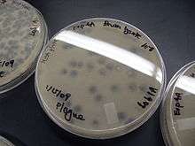
The appearance of the plaque depends on the host strain, virus and the conditions. Highly virulent or lytic strains give clear plaques, while strains that only kill a fraction of their hosts (due to partial resistance/lysogeny) or only reduce the rate of cell growth give turbid plaques. Some partially lysogenic phages give bull's-eye plaques with spots or rings of growth in the middle of clear regions of complete lysis.
Non-viral spontaneous hole formation in cell culture (e.g. LLC-PK1, or the human gingival epithelial cell culture model, Gie-3B11) is called opiplasi (Greek; opi=hole; plasi=formation). These holes can grow to several millimeter in size. Spontaneous appearance of these holes can be induced and accelerated by proinflammatory cytokines such as Tumor Necrosis Factor-alpha [3] .
See also
References
- Finter, N. B (1969-10-01). "Dye Uptake Methods for Assessing Viral Cytopathogenicity and Their Application to Interferon Assays". Journal of General Virology. 5 (3): 419–427. doi:10.1099/0022-1317-5-3-419. ISSN 0022-1317. Retrieved 2012-03-26.
- Marvin, D. A.; Hohn, B. (1969). "Filamentous bacterial viruses". Bacteriological Reviews. 33 (2): 172–209. PMC 378320. PMID 4979697.
- Rybakovsky, E., Buleza, N.B., Hoxha. K., DiGuilio, K.M., McCluskey, E.S., Friday, C.L., Callaghan, P.J., Moskalenko, D.V., Zuo, B., Thomas, S., and Mullin, J.M. (2019). Spontaneous and cytokine-induced hole formation in epithelial cell layers: Implications for barrier function studies with the gingival cell culture, Gie-3B11, and other epithelial models. Trends in Cell and Molecular Biology 13: 99-114.
External links
- An image of Bacteriophage plaques in agar