Tubercle (bone)
In the human skeleton and that of at least other mammals, a tubercle, tuberosity or apophysis is a protrusion or eminence that serves as an attachment for skeletal muscles. The muscles attach by tendons, where the enthesis is the connective tissue between the tendon and bone.[1] A tuberosity is generally a larger tubercle.
Main tubercles
The humerus has two tubercles, the greater tubercle and the lesser tubercle. These are situated at the proximal end of the bone, that is the end that connects with the scapula. The greater/lesser tubercule is located from the top of the acromion laterally and inferiorly. The radius has two, the radial tuberosity and Lister's tubercle.
On a rib, tubercle is an eminence on the back surface, at the junction between the neck and the body of the rib. It consists of an articular and a non-articular area. The lower and more medial articular area is a small oval surface for articulation with the transverse process of the lower of the two vertebrae which gives attachment to the head. The higher, non-articular area is a rough elevation which gives attachment to the ligament of the tubercle. The tubercle is much more prominent in the upper ribs than in the lower ribs.
The most prominent tubercle of the tibia, a leg bone which is more commonly known as the shinbone or shankbone, is the tibial tuberosity. The tibial tuberosity is located on the tibia's anterior surface, distal to the medial condyle. It creates a bony prominence just below the patella, and can be easily located with the fingers. It creates an attachment point for the ligamentum patellae, or patellar ligament. Other tubercles of the tibia include the medial intercondylar tubercle, the lateral intercondylar tubercle, and Gerdy's tubercle.
A trochanter is one of up to three tubercles of the femur:
- Greater trochanter
- Lesser trochanter
- Third trochanter, which is occasionally present
- Fifth metatarsal
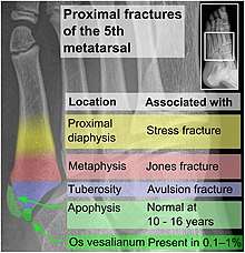
- Proximal diaphysis, typically stress fracture.[2][3]
- Metaphysis: Jones fracture[4]
-Tuberosity: Pseudo-Jones fracture[5] (avulsion fracture).[6]
Normal anatomy:
- Apophysis: Normal at 10 - 16 years.[7]
- Os vesalianum, an accessory bone.[8]
In the fifth metatarsal bone, the most proximal part of the bone is termed the "tuberosity", and the secondary ossification center that is normally present thereon in children is termed the "apophysis".
Related diseases and conditions
- Fractures
The main type of fracture affecting tubercles is avulsion fracture, by pulling the attached tendon.
- Apophysitis
Apophysitis is an inflammation of a tubercle. It mainly affects growing children, with overuse of the affected tubercle.[9][10][11] Examples include:
- Osgood–Schlatter disease (apophysitis of the tibial tubercle)[9]
- Sever's disease (apophysitis of the posterior tubercle of the heel)[10][11]
- Sinding-Larsen and Johansson syndrome (apophysitis of the inferior pole of the patella)[12]
Enthesitis
Enthesitis is an anatomically close but separate condition, wherein there is inflammation of the entheses, the sites where tendons or ligaments insert into the bone.[13][14] It is associated with HLA B27 arthropathies such as ankylosing spondylitis, psoriatic arthritis, and reactive arthritis.[15][16]
References
- "enthesis". Medcyclopaedia. GE. Archived from the original on 2012-02-05.
- Bica D, Sprouse RA, Armen J (2016). "Diagnosis and Management of Common Foot Fractures". Am Fam Physician. 93 (3): 183–91. PMID 26926612.CS1 maint: multiple names: authors list (link)
- "5th Metatarsal". Emergency Care Institute, New South Wales. 2017-09-19.
- "Toe and Forefoot Fractures". OrthoInfo - AAOS. June 2016. Archived from the original on 16 October 2017. Retrieved 15 October 2017.
- Robert Silbergleit. "Foot Fracture". Medscape.com. Retrieved 19 October 2011.
- Robert Silbergleit. "Foot Fracture". Medscape.com. Retrieved October 19, 2011.
- Deniz, G.; Kose, O.; Guneri, B.; Duygun, F. (2014). "Traction apophysitis of the fifth metatarsal base in a child: Iselin's disease". Case Reports. 2014 (may14 4): bcr2014204687–bcr2014204687. doi:10.1136/bcr-2014-204687. ISSN 1757-790X.
- Nwawka, O. Kenechi; Hayashi, Daichi; Diaz, Luis E.; Goud, Ajay R.; Arndt, William F.; Roemer, Frank W.; Malguria, Nagina; Guermazi, Ali (2013). "Sesamoids and accessory ossicles of the foot: anatomical variability and related pathology". Insights into Imaging. 4 (5): 581–593. doi:10.1007/s13244-013-0277-1. ISSN 1869-4101. PMC 3781258.
- "OrthoKids - Osgood-Schlatter's Disease".
- "Sever's Disease". Kidshealth.org. Retrieved 2014-04-29.
- Hendrix CL (2005). "Calcaneal apophysitis (Sever disease)". Clinics in Podiatric Medicine and Surgery. 22 (1): 55–62, vi. doi:10.1016/j.cpm.2004.08.011. PMID 15555843.
- "Sinding-Larsen-Johansson Syndrome - Knee & Sports - Orthobullets". www.orthobullets.com. Retrieved 2019-08-05.
- Maria Antonietta D'Agostino, MD; Ignazio Olivieri, MD (June 2006). "Enthesitis". Best Practice & Research Clinical Rheumatology. Clinical Rheumatology. 20 (3): 473–86. doi:10.1016/j.berh.2006.03.007.
- The Free Dictionary (2009). "Enthesitis". Retrieved 2010-11-27.
- Schett, G; Lories, RJ; D'Agostino, MA; Elewaut, D; Kirkham, B; Soriano, ER; McGonagle, D (November 2017). "Enthesitis: from pathophysiology to treatment". Nature Reviews Rheumatology (Review). 13 (12): 731–41. doi:10.1038/nrrheum.2017.188. PMID 29158573.
- Schmitt, SK (June 2017). "Reactive Arthritis". Infectious Disease Clinics of North America (Review). 31 (2): 265–77. doi:10.1016/j.idc.2017.01.002. PMID 28292540.