Transcriptional regulation
In molecular biology and genetics, transcriptional regulation is the means by which a cell regulates the conversion of DNA to RNA (transcription), thereby orchestrating gene activity. A single gene can be regulated in a range of ways, from altering the number of copies of RNA that are transcribed, to the temporal control of when the gene is transcribed. This control allows the cell or organism to respond to a variety of intra- and extracellular signals and thus mount a response. Some examples of this include producing the mRNA that encode enzymes to adapt to a change in a food source, producing the gene products involved in cell cycle specific activities, and producing the gene products responsible for cellular differentiation in multicellular eukaryotes, as studied in evolutionary developmental biology.
| Transcription regulation glossary |
|---|
| • transcriptional regulation – controlling the rate of gene transcription for example by helping or hindering RNA polymerase binding to DNA |
| • transcription – the process of making RNA from a DNA template by RNA polymerase |
| • transcription factor – a substance, such as a protein, that contributes to the cause of a specific biochemical reaction or bodily process |
| • promoter – a region of DNA that initiates transcription of a particular gene |
| • Sigma factor – specialized bacterial co-factors that complex with RNA Polymerase and encode sequence specificity |
| • coactivator – a protein that works with transcription factors to increase the rate of gene transcription |
| • corepressor – a protein that works with transcription factors to decrease the rate of gene transcription |
The regulation of transcription is a vital process in all living organisms. It is orchestrated by transcription factors and other proteins working in concert to finely tune the amount of RNA being produced through a variety of mechanisms. Prokaryotic organisms and eukaryotic organisms have very different strategies of accomplishing control over transcription, but some important features remain conserved between the two. Most importantly is the idea of combinatorial control, which is that any given gene is likely controlled by a specific combination of factors to control transcription. In a hypothetical example, the factors A and B might regulate a distinct set of genes from the combination of factors A and C. This combinatorial nature extends to complexes of far more than two proteins, and allows a very small subset (less than 10%) of the genome to control the transcriptional program of the entire cell.
In prokaryotes

Much of the early understanding of transcription came from prokaryotic organisms,[2] although the extent and complexity of transcriptional regulation is greater in eukaryotes. Prokaryotic transcription is governed by three main sequence elements:
- Promoters are elements of DNA that may bind RNA polymerase and other proteins for the successful initiation of transcription directly upstream of the gene.
- Operators recognize repressor proteins that bind to a stretch of DNA and inhibit the transcription of the gene.
- Positive control elements that bind to DNA and incite higher levels of transcription.[3]
While these means of transcriptional regulation also exist in eukaryotes, the transcriptional landscape is significantly more complicated both by the number of proteins involved as well as by the presence of introns and the packaging of DNA into histones.
The transcription of a basic prokaryotic gene is dependent on the strength of its promoter and the presence of activators or repressors. In the absence of other regulatory elements, a promoter's sequence-based affinity for RNA polymerases varies, which results in the production of different amounts of transcript. The variable affinity of RNA polymerase for different promoter sequences is related to regions of consensus sequence upstream of the transcription start site. The more nucleotides of a promoter that agree with the consensus sequence, the stronger the affinity of the promoter for RNA Polymerase likely is.[4]

In the absence of other regulatory elements, the default state of a prokaryotic transcript is to be in the “on” configuration, resulting in the production of some amount of transcript. This means that transcriptional regulation in the form of protein repressors and positive control elements can either increase or decrease transcription. Repressors often physically occupy the promoter location, occluding RNA polymerase from binding. Alternatively a repressor and polymerase may bind to the DNA at the same time with a physical interaction between the repressor preventing the opening of the DNA for access to the minus strand for transcription. This strategy of control is distinct from eukaryotic transcription, whose basal state is to be off and where co-factors required for transcription initiation are highly gene dependent.[8]
Sigma factors are specialized bacterial proteins that bind to RNA polymerases and orchestrate transcription initiation. Sigma factors act as mediators of sequence-specific transcription, such that a single sigma factor can be used for transcription of all housekeeping genes or a suite of genes the cell wishes to express in response to some external stimuli such as stress.[9]
In eukaryotes
The added complexity of generating a eukaryotic cell carries with it an increase in the complexity of transcriptional regulation. Eukaryotes have three RNA polymerases, known as Pol I, Pol II, and Pol III. Each polymerase has specific targets and activities, and is regulated by independent mechanisms. There are a number of additional mechanisms through which polymerase activity can be controlled. These mechanisms can be generally grouped into three main areas:
- Control over polymerase access to the gene. This is perhaps the broadest of the three control mechanisms. This includes the functions of histone remodeling enzymes, transcription factors, enhancers and repressors, and many other complexes
- Productive elongation of the RNA transcript. Once polymerase is bound to a promoter, it requires another set of factors to allow it to escape the promoter complex and begin successfully transcribing RNA.
- Termination of the polymerase. A number of factors which have been found to control how and when termination occurs, which will dictate the fate of the RNA transcript.
All three of these systems work in concert to integrate signals from the cell and change the transcriptional program accordingly.
While in prokaryotic systems the basal transcription state can be thought of as nonrestrictive (that is, “on” in the absence of modifying factors), eukaryotes have a restrictive basal state which requires the recruitment of other factors in order to generate RNA transcripts. This difference is largely due to the compaction of the eukaryotic genome by winding DNA around histones to form higher order structures. This compaction makes the gene promoter inaccessible without the assistance of other factors in the nucleus, and thus chromatin structure is a common site of regulation. Similar to the sigma factors in prokaryotes, the general transcription factors (GTFs) are a set of factors in eukaryotes that are required for all transcription events. These factors are responsible for stabilizing binding interactions and opening the DNA helix to allow the RNA polymerase to access the template, but generally lack specificity for different promoter sites.[10] A large part of gene regulation occurs through transcription factors that either recruit or inhibit the binding of the general transcription machinery and/or the polymerase. This can be accomplished through close interactions with core promoter elements, or through the long distance enhancer elements.
Once a polymerase is successfully bound to a DNA template, it often requires the assistance of other proteins in order to leave the stable promoter complex and begin elongating the nascent RNA strand. This process is called promoter escape, and is another step at which regulatory elements can act to accelerate or slow the transcription process. Similarly, protein and nucleic acid factors can associate with the elongation complex and modulate the rate at which the polymerase moves along the DNA template.
At the level of chromatin state
In eukaryotes, genomic DNA is highly compacted in order to be able to fit it into the nucleus. This is accomplished by winding the DNA around protein octamers called histones, which has consequences for the physical accessibility of parts of the genome at any given time. Significant portions are silenced through histone modifications, and thus are inaccessible to the polymerases or their cofactors. The highest level of transcription regulation occurs through the rearrangement of histones in order to expose or sequester genes, because these processes have the ability to render entire regions of a chromosome inaccessible such as what occurs in imprinting.
Histone rearrangement is facilitated by post-translational modifications to the tails of the core histones. A wide variety of modifications can be made by enzymes such as the histone acetyltransferases (HATs), histone methyltransferases (HMTs), and histone deacetylases (HDACs), among others. These enzymes can add or remove covalent modifications such as methyl groups, acetyl groups, phosphates, and ubiquitin. Histone modifications serve to recruit other proteins which can either increase the compaction of the chromatin and sequester promoter elements, or to increase the spacing between histones and allow the association of transcription factors or polymerase on open DNA.[11] For example, H3K27 trimethylation by the polycomb complex PRC2 causes chromosomal compaction and gene silencing.[12] These histone modifications may be created by the cell, or inherited in an epigenetic fashion from a parent.
At the level of cytosine methylation
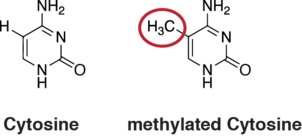
5-Methylcytosine is a methylated form of the DNA base cytosine (C) that regulates gene transcription in mammals.[13] Methylated cytosines primarily occur in dinucleotide sequences where cytosine is followed by a guanine, a CpG site. The total number of CpG sites in the human genome is approximately 28 million.[14] and generally about 70% of all CpG sites have a methylated cytosine.[15] Methylation of CpGs in a promoter region of a gene represses transcription[16] while methylation of CpGs in the body of a gene increases expression.[17] TET enzymes play a central role in demethylation of methylated cytosines. Demethylation of CpGs in a gene promoter by TET enzyme activity increases transcription of the gene.[18]
Through transcription factors and enhancers
Transcription factors
Transcription factors are proteins that bind to specific DNA sequences in order to regulate the expression of a given gene. There are approximately 1,400 transcription factors in the human genome and they constitute about 6% of all human protein coding genes.[19] The power of transcription factors resides in their ability to activate and/or repress wide repertoires of downstream target genes. The fact that these transcription factors work in a combinatorial fashion means that only a small subset of an organism's genome encodes transcription factors. Transcription factors function through a wide variety of mechanisms. In one mechanism, CpG methylation influences binding of most transcription factors to DNA—in some cases negatively and in others positively. [20] In addition, often they are at the end of a signal transduction pathway that functions to change something about the factor, like its subcellular localization or its activity. Post-translational modifications to transcription factors located in the cytosol can cause them to translocate to the nucleus where they can interact with their corresponding enhancers. Other transcription factors are already in the nucleus, and are modified to enable the interaction with partner transcription factors. Some post-translational modifications known to regulate the functional state of transcription factors are phosphorylation, acetylation, SUMOylation and ubiquitylation. Transcription factors can be divided in two main categories: activators and repressors. While activators can interact directly or indirectly with the core machinery of transcription through enhancer binding, repressors predominantly recruit co-repressor complexes leading to transcriptional repression by chromatin condensation of enhancer regions. It may also happen that a repressor may function by allosteric competition against a determined activator to repress gene expression: overlapping DNA-binding motifs for both activators and repressors induce a physical competition to occupy the site of binding. If the repressor has a higher affinity for its motif than the activator, transcription would be effectively blocked in the presence of the repressor. Tight regulatory control is achieved by the highly dynamic nature of transcription factors. Again, many different mechanisms exist to control whether a transcription factor is active. These mechanisms include control over protein localization or control over whether the protein can bind DNA.[21] An example of this is the protein HSF1, which remains bound to Hsp70 in the cytosol and is only translocated into the nucleus upon cellular stress such as heat shock. Thus the genes under the control of this transcription factor will remain untranscribed unless the cell is subjected to stress.[22]
Enhancers
Enhancers or cis-regulatory modules/elements (CRM/CRE) are non-coding DNA sequences containing multiple activator and repressor binding sites. Enhancers range from 200 bp to 1 kb in length and can be either proximal, 5’ upstream to the promoter or within the first intron of the regulated gene, or distal, in introns of neighboring genes or intergenic regions far away from the locus. Through DNA looping, active enhancers contact the promoter dependently of the core DNA binding motif promoter specificity.[23] Promoter-enhancer dichotomy provides the basis for the functional interaction between transcription factors and transcriptional core machinery to trigger RNA Pol II escape from the promoter. Whereas one could think that there is a 1:1 enhancer-promoter ratio, studies of the human genome predict that an active promoter interacts with 4 to 5 enhancers. Similarly, enhancers can regulate more than one gene without linkage restriction and are said to “skip” neighboring genes to regulate more distant ones. Even though infrequent, transcriptional regulation can involve elements located in a chromosome different to one where the promoter resides. Proximal enhancers or promoters of neighboring genes can serve as platforms to recruit more distal elements.[24]
Regulatory landscape
Transcriptional initiation, termination and regulation are mediated by “DNA looping” which brings together promoters, enhancers, transcription factors and RNA processing factors to accurately regulate gene expression.[25] Chromosome conformation capture (3C) and more recently Hi-C techniques provided evidence that active chromatin regions are “compacted” in nuclear domains or bodies where transcriptional regulation is enhanced.[26] The configuration of the genome is essential for enhancer-promoter proximity. Cell-fate decisions are mediated upon highly dynamic genomic reorganizations at interphase to modularly switch on or off entire gene regulatory networks through short to long range chromatin rearrangements.[27] Related studies demonstrate that metazoan genomes are partitioned in structural and functional units around a megabase long called Topological association domains (TADs) containing dozens of genes regulated by hundreds of enhancers distributed within large genomic regions containing only non-coding sequences. The function of TADs is to regroup enhancers and promoters interacting together within a single large functional domain instead of having them spread in different TADs.[28] However, studies of mouse development point out that two adjacent TADs may regulate the same gene cluster. The most relevant study on limb evolution shows that the TAD at the 5’ of the HoxD gene cluster in tetrapod genomes drives its expression in the distal limb bud embryos, giving rise to the hand, while the one located at 3’ side does it in the proximal limb bud, giving rise to the arm.[29] Still, it is not known whether TADs are an adaptive strategy to enhance regulatory interactions or an effect of the constrains on these same interactions. TAD boundaries are often composed by housekeeping genes, tRNAs, other highly expressed sequences and Short Interspersed Elements (SINE). While these genes may take advantage of their border position to be ubiquitously expressed, they are not directly linked with TAD edge formation. The specific molecules identified at boundaries of TADs are called insulators or architectural proteins because they not only block enhancer leaky expression but also ensure an accurate compartmentalization of cis-regulatory inputs to the targeted promoter. These insulators are DNA-binding proteins like CTCF and TFIIIC that help recruiting structural partners such as cohesins and condensins. The localization and binding of architectural proteins to their corresponding binding sites is regulated by post-translational modifications.[30] DNA binding motifs recognized by architectural proteins are either of high occupancy and at around a megabase of each other or of low occupancy and inside TADs. High occupancy sites are usually conserved and static while intra-TADs sites are dynamic according to the state of the cell therefore TADs themselves are compartmentalized in subdomains that can be called subTADs from few kb up to a TAD long (19). When architectural binding sites are at less than 100 kb from each other, Mediator proteins are the architectural proteins cooperate with cohesin. For subTADs larger than 100 kb and TAD boundaries, CTCF is the typical insulator found to interact with cohesion.[31]
Of the pre-initiation complex and promoter escape
In eukaryotes, ribosomal rRNA and the tRNAs involved in translation are controlled by RNA polymerase I (Pol I) and RNA polymerase III (Pol III) . RNA Polymerase II (Pol II) is responsible for the production of messenger RNA (mRNA) within the cell. Particularly for Pol II, much of the regulatory checkpoints in the transcription process occur in the assembly and escape of the pre-initiation complex. A gene-specific combination of transcription factors will recruit TFIID and/or TFIIA to the core promoter, followed by the association of TFIIB, creating a stable complex onto which the rest of the General Transcription Factors (GTFs) can assemble.[32] This complex is relatively stable, and can undergo multiple rounds of transcription initiation.[33] After the binding of TFIIB and TFIID, Pol II the rest of the GTFs can assemble. This assembly is marked by the post-translational modification (typically phosphorylation) of the C-terminal domain (CTD) of Pol II through a number of kinases.[34] The CTD is a large, unstructured domain extending from the RbpI subunit of Pol II, and consists of many repeats of the heptad sequence YSPTSPS. TFIIH, the helicase that remains associated with Pol II throughout transcription, also contains a subunit with kinase activity which will phosphorylate the serines 5 in the heptad sequence. Similarly, both CDK8 (a subunit of the massive multiprotein Mediator complex) and CDK9 (a subunit of the p-TEFb elongation factor), have kinase activity towards other residues on the CTD.[35] These phosphorylation events promote the transcription process and serve as sites of recruitment for mRNA processing machinery. All three of these kinases respond to upstream signals, and failure to phosphorylate the CTD can lead to a stalled polymerase at the promoter.
In cancer
In vertebrates, the majority of gene promoters contain a CpG island with numerous CpG sites.[36] When many of a gene's promoter CpG sites are methylated the gene becomes silenced.[37] Colorectal cancers typically have 3 to 6 driver mutations and 33 to 66 hitchhiker or passenger mutations.[38] However, transcriptional silencing may be of more importance than mutation in causing progression to cancer. For example, in colorectal cancers about 600 to 800 genes are transcriptionally silenced by CpG island methylation (see regulation of transcription in cancer). Transcriptional repression in cancer can also occur by other epigenetic mechanisms, such as altered expression of microRNAs.[39] In breast cancer, transcriptional repression of BRCA1 may occur more frequently by over-expressed microRNA-182 than by hypermethylation of the BRCA1 promoter (see Low expression of BRCA1 in breast and ovarian cancers).
References
- Madigan, Michael T. Brock Biology of Microorganisms, 15e. Pearson. p. 178. ISBN 9780134602295.
- JACOB F, MONOD J (June 1961). "Genetic regulatory mechanisms in the synthesis of proteins". J. Mol. Biol. 3 (3): 318–56. doi:10.1016/s0022-2836(61)80072-7. PMID 13718526.
- Englesberg E, Irr J, Power J, Lee N (October 1965). "Positive control of enzyme synthesis by gene C in the L-arabinose system". J. Bacteriol. 90 (4): 946–57. PMC 315760. PMID 5321403.
- Busby S, Ebright RH (December 1994). "Promoter structure, promoter recognition, and transcription activation in prokaryotes". Cell. 79 (5): 743–6. doi:10.1016/0092-8674(94)90063-9. PMID 8001112.
- "malE - Maltose-binding periplasmic protein precursor - Escherichia coli (strain K12) - malE gene & protein". www.uniprot.org. Retrieved 20 November 2017.
- "malF - Maltose transport system permease protein MalF - Escherichia coli (strain K12) - malF gene & protein". www.uniprot.org. Retrieved 20 November 2017.
- "malG - Maltose transport system permease protein MalG - Escherichia coli (strain K12) - malG gene & protein". www.uniprot.org. Retrieved 20 November 2017.
- Payankaulam S, Li LM, Arnosti DN (September 2010). "Transcriptional repression: conserved and evolved features". Curr. Biol. 20 (17): R764–71. doi:10.1016/j.cub.2010.06.037. PMC 3033598. PMID 20833321.
- Gruber TM, Gross CA (2003). "Multiple sigma subunits and the partitioning of bacterial transcription space". Annu. Rev. Microbiol. 57: 441–66. doi:10.1146/annurev.micro.57.030502.090913. PMID 14527287.
- Struhl K (July 1999). "Fundamentally different logic of gene regulation in eukaryotes and prokaryotes". Cell. 98 (1): 1–4. doi:10.1016/S0092-8674(00)80599-1. PMID 10412974.
- Calo E, Wysocka J (March 2013). "Modification of enhancer chromatin: what, how, and why?". Mol. Cell. 49 (5): 825–37. doi:10.1016/j.molcel.2013.01.038. PMC 3857148. PMID 23473601.
- de Napoles M, Mermoud JE, Wakao R, Tang YA, Endoh M, Appanah R, Nesterova TB, Silva J, Otte AP, Vidal M, Koseki H, Brockdorff N (November 2004). "Polycomb group proteins Ring1A/B link ubiquitylation of histone H2A to heritable gene silencing and X inactivation". Dev. Cell. 7 (5): 663–76. doi:10.1016/j.devcel.2004.10.005. PMID 15525528.
- Turek-Plewa J, Jagodziński PP (2005). "The role of mammalian DNA methyltransferases in the regulation of gene expression". Cell. Mol. Biol. Lett. 10 (4): 631–47. PMID 16341272.
- Lövkvist C, Dodd IB, Sneppen K, Haerter JO (June 2016). "DNA methylation in human epigenomes depends on local topology of CpG sites". Nucleic Acids Research. 44 (11): 5123–32. doi:10.1093/nar/gkw124. PMC 4914085. PMID 26932361.
- Jabbari K, Bernardi G (May 2004). "Cytosine methylation and CpG, TpG (CpA) and TpA frequencies". Gene. 333: 143–9. doi:10.1016/j.gene.2004.02.043. PMID 15177689.
- Weber M, Hellmann I, Stadler MB, Ramos L, Pääbo S, Rebhan M, Schübeler D (April 2007). "Distribution, silencing potential and evolutionary impact of promoter DNA methylation in the human genome". Nat. Genet. 39 (4): 457–66. doi:10.1038/ng1990. PMID 17334365.
- Yang X, Han H, De Carvalho DD, Lay FD, Jones PA, Liang G (October 2014). "Gene body methylation can alter gene expression and is a therapeutic target in cancer". Cancer Cell. 26 (4): 577–90. doi:10.1016/j.ccr.2014.07.028. PMC 4224113. PMID 25263941.
- Maeder ML, Angstman JF, Richardson ME, Linder SJ, Cascio VM, Tsai SQ, Ho QH, Sander JD, Reyon D, Bernstein BE, Costello JF, Wilkinson MF, Joung JK (December 2013). "Targeted DNA demethylation and activation of endogenous genes using programmable TALE-TET1 fusion proteins". Nat. Biotechnol. 31 (12): 1137–42. doi:10.1038/nbt.2726. PMC 3858462. PMID 24108092.
- Vaquerizas JM, Kummerfeld SK, Teichmann SA, Luscombe NM (April 2009). "A census of human transcription factors: function, expression and evolution". Nat. Rev. Genet. 10 (4): 252–63. doi:10.1038/nrg2538. PMID 19274049.
- Yin Y, Morgunova E, Jolma A, Kaasinen E, Sahu B, Khund-Sayeed S, Das PK, Kivioja T, Dave K, Zhong F, Nitta KR, Taipale M, Popov A, Ginno PA, Domcke S, Yan J, Schübeler D, Vinson C, Taipale J (May 2017). "Impact of cytosine methylation on DNA binding specificities of human transcription factors". Science. 356 (6337). doi:10.1126/science.aaj2239. PMID 28473536.
- Whiteside ST, Goodbourn S (April 1993). "Signal transduction and nuclear targeting: regulation of transcription factor activity by subcellular localisation". J. Cell Sci. 104 ( Pt 4): 949–55. PMID 8314906.
- Vihervaara A, Sistonen L (January 2014). "HSF1 at a glance". J. Cell Sci. 127 (Pt 2): 261–6. doi:10.1242/jcs.132605. PMID 24421309.
- Levine M (September 2010). "Transcriptional enhancers in animal development and evolution". Curr. Biol. 20 (17): R754–63. doi:10.1016/j.cub.2010.06.070. PMC 4280268. PMID 20833320.
- van Arensbergen J, van Steensel B, Bussemaker HJ (November 2014). "In search of the determinants of enhancer-promoter interaction specificity". Trends Cell Biol. 24 (11): 695–702. doi:10.1016/j.tcb.2014.07.004. PMC 4252644. PMID 25160912.
- Mercer TR, Mattick JS (July 2013). "Understanding the regulatory and transcriptional complexity of the genome through structure". Genome Res. 23 (7): 1081–8. doi:10.1101/gr.156612.113. PMC 3698501. PMID 23817049.
- Dekker J, Marti-Renom MA, Mirny LA (June 2013). "Exploring the three-dimensional organization of genomes: interpreting chromatin interaction data". Nat. Rev. Genet. 14 (6): 390–403. doi:10.1038/nrg3454. PMC 3874835. PMID 23657480.
- Gómez-Díaz E, Corces VG (November 2014). "Architectural proteins: regulators of 3D genome organization in cell fate". Trends Cell Biol. 24 (11): 703–11. doi:10.1016/j.tcb.2014.08.003. PMC 4254322. PMID 25218583.
- Smallwood A, Ren B (June 2013). "Genome organization and long-range regulation of gene expression by enhancers". Curr. Opin. Cell Biol. 25 (3): 387–94. doi:10.1016/j.ceb.2013.02.005. PMC 4180870. PMID 23465541.
- Woltering JM, Noordermeer D, Leleu M, Duboule D (January 2014). "Conservation and divergence of regulatory strategies at Hox Loci and the origin of tetrapod digits". PLoS Biol. 12 (1): e1001773. doi:10.1371/journal.pbio.1001773. PMC 3897358. PMID 24465181.
- Wang H, Maurano MT, Qu H, Varley KE, Gertz J, Pauli F, Lee K, Canfield T, Weaver M, Sandstrom R, Thurman RE, Kaul R, Myers RM, Stamatoyannopoulos JA (September 2012). "Widespread plasticity in CTCF occupancy linked to DNA methylation". Genome Res. 22 (9): 1680–8. doi:10.1101/gr.136101.111. PMC 3431485. PMID 22955980.
- Phillips-Cremins JE, Sauria ME, Sanyal A, Gerasimova TI, Lajoie BR, Bell JS, et al. (June 2013). "Architectural protein subclasses shape 3D organization of genomes during lineage commitment". Cell. 153 (6): 1281–95. doi:10.1016/j.cell.2013.04.053. PMC 3712340. PMID 23706625.
- Thomas MC, Chiang CM (2006). "The general transcription machinery and general cofactors". Crit. Rev. Biochem. Mol. Biol. 41 (3): 105–78. CiteSeerX 10.1.1.376.5724. doi:10.1080/10409230600648736. PMID 16858867.
- Voet, Donald Voet, Judith G. (2011). Biochemistry (4th ed.). Hoboken, NJ: John Wiley & Sons. ISBN 978-0470917459.
- Napolitano G, Lania L, Majello B (May 2014). "RNA polymerase II CTD modifications: how many tales from a single tail". J. Cell. Physiol. 229 (5): 538–44. doi:10.1002/jcp.24483. PMID 24122273.
- Chapman RD, Conrad M, Eick D (September 2005). "Role of the mammalian RNA polymerase II C-terminal domain (CTD) nonconsensus repeats in CTD stability and cell proliferation". Mol. Cell. Biol. 25 (17): 7665–74. doi:10.1128/MCB.25.17.7665-7674.2005. PMC 1190292. PMID 16107713.
- Saxonov S, Berg P, Brutlag DL (2006). "A genome-wide analysis of CpG dinucleotides in the human genome distinguishes two distinct classes of promoters". Proc. Natl. Acad. Sci. U.S.A. 103 (5): 1412–7. doi:10.1073/pnas.0510310103. PMC 1345710. PMID 16432200.
- Bird A (2002). "DNA methylation patterns and epigenetic memory". Genes Dev. 16 (1): 6–21. doi:10.1101/gad.947102. PMID 11782440.
- Vogelstein B, Papadopoulos N, Velculescu VE, Zhou S, Diaz LA, Kinzler KW (2013). "Cancer genome landscapes". Science. 339 (6127): 1546–58. doi:10.1126/science.1235122. PMC 3749880. PMID 23539594.
- Tessitore A, Cicciarelli G, Del Vecchio F, Gaggiano A, Verzella D, Fischietti M, Vecchiotti D, Capece D, Zazzeroni F, Alesse E (2014). "MicroRNAs in the DNA Damage/Repair Network and Cancer". Int J Genom. 2014: 1–10. doi:10.1155/2014/820248. PMC 3926391. PMID 24616890.