PMEL (gene)
Melanocyte protein PMEL also known as premelanosome protein (PMEL) or silver locus protein homolog (SILV) is a protein that in humans is encoded by the PMEL gene.[5][6] Its gene product may be referred to as PMEL, silver, ME20, gp100 or Pmel17.


Structure and function
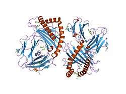
PMEL is a 100 kDa type I transmembrane glycoprotein that is expressed primarily in pigment cells of the skin and eye. The transmembrane form of PMEL is modified in the secretory pathway by elaboration of N-linked oligosaccharides and addition and modification of O-linked oligosaccharides. It is then targeted to precursors of the pigment organelle, the melanosome, where it is proteolytically processed to several small fragments. Some of these fragments form non-pathological amyloid that assemble into sheets and form the striated pattern that underlies melanosomal ultrastructure. PMEL cleavage is mediated by several proteases including a proprotein convertase of the furin family, a "sheddase" that might include members of the a disintegrin and metalloproteinase (ADAM) family, and additional proteases in melanosomes or their precursors. After the amyloidogenic region is cleaved, the small remaining integral membrane fragment is digested by γ-secretase.
The expression of the PMEL gene is regulated by the microphthalmia-associated transcription factor (MITF).[7][8]
References
- GRCh38: Ensembl release 89: ENSG00000185664 - Ensembl, May 2017
- GRCm38: Ensembl release 89: ENSMUSG00000025359 - Ensembl, May 2017
- "Human PubMed Reference:". National Center for Biotechnology Information, U.S. National Library of Medicine.
- "Mouse PubMed Reference:". National Center for Biotechnology Information, U.S. National Library of Medicine.
- Kim KK, Youn BS, Heng HH, Shi XM, Tsui LC, Lee ZH, Pickard RT, Kwon BS (Oct 1996). "Genomic organization and FISH mapping of human Pmel 17, the putative silver locus". Pigment Cell Res. 9 (1): 42–8. doi:10.1111/j.1600-0749.1996.tb00085.x. PMID 8739560.
- "Entrez Gene: SILV silver homolog (mouse)".
- Du J, Miller AJ, Widlund HR, Horstmann MA, Ramaswamy S, Fisher DE (2003). "MLANA/MART1 and SILV/PMEL17/GP100 are transcriptionally regulated by MITF in melanocytes and melanoma". Am. J. Pathol. 163 (1): 333–43. doi:10.1016/S0002-9440(10)63657-7. PMC 1868174. PMID 12819038.
- Hoek KS, Schlegel NC, Eichhoff OM, Widmer DS, Praetorius C, Einarsson SO, Valgeirsdottir S, Bergsteinsdottir K, Schepsky A, Dummer R, Steingrimsson E (2008). "Novel MITF targets identified using a two-step DNA microarray strategy". Pigment Cell Melanoma Res. 21 (6): 665–76. doi:10.1111/j.1755-148X.2008.00505.x. PMID 19067971.
Further reading
- Aplan PD, Lombardi DP, Kirsch IR (1991). "Structural characterization of SIL, a gene frequently disrupted in T-cell acute lymphoblastic leukemia". Mol. Cell. Biol. 11 (11): 5462–9. doi:10.1128/mcb.11.11.5462. PMC 361915. PMID 1922059.
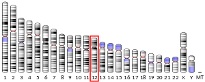
- Kwon BS, Chintamaneni C, Kozak CA, et al. (1991). "A melanocyte-specific gene, Pmel 17, maps near the silver coat color locus on mouse chromosome 10 and is in a syntenic region on human chromosome 12". Proc. Natl. Acad. Sci. U.S.A. 88 (20): 9228–32. doi:10.1073/pnas.88.20.9228. PMC 52687. PMID 1924386.
- Brown L, Cheng JT, Chen Q, et al. (1990). "Site-specific recombination of the tal-1 gene is a common occurrence in human T cell leukemia". EMBO J. 9 (10): 3343–51. doi:10.1002/j.1460-2075.1990.tb07535.x. PMC 552072. PMID 2209547.
- Guillaume van Niel; Ptissam Bergam; Aurelie Di Cicco; et al. (2015). "Apolipoprotein E Regulates Amyloid Formation within Endosomes of Pigment Cells". Cell Reports. 13 (1): 43–51. doi:10.1016/j.celrep.2015.08.057. PMID 26387950.
- Adema GJ, de Boer AJ, Vogel AM, et al. (1994). "Molecular characterization of the melanocyte lineage-specific antigen gp100". J. Biol. Chem. 269 (31): 20126–33. PMID 7519602.
- Kawakami Y, Eliyahu S, Delgado CH, et al. (1994). "Identification of a human melanoma antigen recognized by tumor-infiltrating lymphocytes associated with in vivo tumor rejection". Proc. Natl. Acad. Sci. U.S.A. 91 (14): 6458–62. doi:10.1073/pnas.91.14.6458. PMC 44221. PMID 8022805.
- Maresh GA, Marken JS, Neubauer M, et al. (1994). "Cloning and expression of the gene for the melanoma-associated ME20 antigen". DNA Cell Biol. 13 (2): 87–95. doi:10.1089/dna.1994.13.87. PMID 8179825.
- Bailin T, Lee ST, Spritz RA (1996). "Genomic organization and sequence of D12S53E (Pmel 17), the human homologue of the mouse silver (si) locus". J. Invest. Dermatol. 106 (1): 24–7. doi:10.1111/1523-1747.ep12326976. PMID 8592076.
- Chakraborty AK, Platt JT, Kim KK, et al. (1996). "Polymerization of 5,6-dihydroxyindole-2-carboxylic acid to melanin by the pmel 17/silver locus protein". Eur. J. Biochem. 236 (1): 180–8. doi:10.1111/j.1432-1033.1996.t01-1-00180.x. PMID 8617263.
- Chi DD, Merchant RE, Rand R, et al. (1997). "Molecular detection of tumor-associated antigens shared by human cutaneous melanomas and gliomas". Am. J. Pathol. 150 (6): 2143–52. PMC 1858329. PMID 9176405.
- Wagner SN, Wagner C, Schultewolter T, Goos M (1997). "Analysis of Pmel17/gp100 expression in primary human tissue specimens: implications for melanoma immuno- and gene-therapy". Cancer Immunol. Immunother. 44 (4): 239–47. doi:10.1007/s002620050379. PMID 9222283.
- Curry BJ, Myers K, Hersey P (1998). "Polymerase chain reaction detection of melanoma cells in the circulation: relation to clinical stage, surgical treatment, and recurrence from melanoma". J. Clin. Oncol. 16 (5): 1760–9. doi:10.1200/JCO.1998.16.5.1760. PMID 9586889.
- Lupetti R, Pisarra P, Verrecchia A, et al. (1998). "Translation of a retained intron in tyrosinase-related protein (TRP) 2 mRNA generates a new cytotoxic T lymphocyte (CTL)-defined and shared human melanoma antigen not expressed in normal cells of the melanocytic lineage". J. Exp. Med. 188 (6): 1005–16. doi:10.1084/jem.188.6.1005. PMC 2212536. PMID 9743519.
- Lukowsky A, Bellmann B, Ringk A, et al. (1999). "Detection of melanoma micrometastases in the sentinel lymph node and in nonsentinel nodes by tyrosinase polymerase chain reaction". J. Invest. Dermatol. 113 (4): 554–9. doi:10.1046/j.1523-1747.1999.00719.x. PMID 10504440.
- Raposo G, Tenza D, Murphy DM, et al. (2001). "Distinct protein sorting and localization to premelanosomes, melanosomes, and lysosomes in pigmented melanocytic cells". J. Cell Biol. 152 (4): 809–24. doi:10.1083/jcb.152.4.809. PMC 2195785. PMID 11266471.
- Virador V, Matsunaga N, Matsunaga J, et al. (2002). "Production of melanocyte-specific antibodies to human melanosomal proteins: expression patterns in normal human skin and in cutaneous pigmented lesions". Pigment Cell Res. 14 (4): 289–97. doi:10.1034/j.1600-0749.2001.140410.x. PMID 11549113.
- Berson JF, Harper DC, Tenza D, et al. (2002). "Pmel17 initiates premelanosome morphogenesis within multivesicular bodies". Mol. Biol. Cell. 12 (11): 3451–64. doi:10.1091/mbc.12.11.3451. PMC 60267. PMID 11694580.
- Sensi M, Pellegatta S, Vegetti C, et al. (2003). "Identification of a novel gp100/pMel17 peptide presented by HLA-A*6801 and recognized on human melanoma by cytolytic T cell clones". Tissue Antigens. 59 (4): 273–9. doi:10.1034/j.1399-0039.2002.590404.x. PMID 12135425.
- Strausberg RL, Feingold EA, Grouse LH, et al. (2003). "Generation and initial analysis of more than 15,000 full-length human and mouse cDNA sequences". Proc. Natl. Acad. Sci. U.S.A. 99 (26): 16899–903. doi:10.1073/pnas.242603899. PMC 139241. PMID 12477932.