Lipid peroxidation
Lipid peroxidation is the oxidative degradation of lipids. It is the process in which free radicals "steal" electrons from the lipids in cell membranes, resulting in cell damage. This process proceeds by a free radical chain reaction mechanism. It most often affects polyunsaturated fatty acids, because they contain multiple double bonds in between which lie methylene bridges (-CH2-) that possess especially reactive hydrogen atoms. As with any radical reaction, the reaction consists of three major steps: initiation, propagation, and termination. The chemical products of this oxidation are known as lipid peroxides or lipid oxidation products (LOPs).
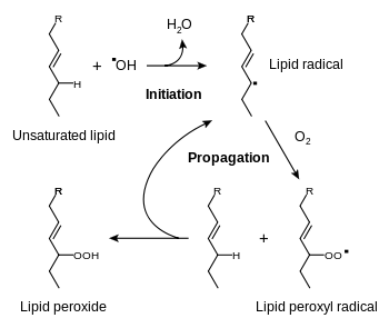
Initiation
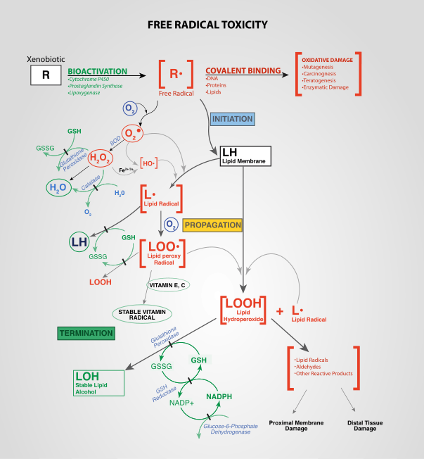
Initiation is the step in which a fatty acid radical is produced. The most notable initiators in living cells are reactive oxygen species (ROS), such as OH· and HOO·, which combines with a hydrogen atom to make water and a fatty acid radical.
Propagation
The fatty acid radical is not a very stable molecule, so it reacts readily with molecular oxygen, thereby creating a peroxyl-fatty acid radical. This radical is also an unstable species that reacts with another free fatty acid, producing a different fatty acid radical and a lipid peroxide, or a cyclic peroxide if it had reacted with itself. This cycle continues, as the new fatty acid radical reacts in the same way.
Termination
When a radical reacts with a non-radical, it always produces another radical, which is why the process is called a "chain reaction mechanism". The radical reaction stops when two radicals react and produce a non-radical species. This happens only when the concentration of radical species is high enough for there to be a high probability of collision of two radicals. Living organisms have different molecules that speed up termination by neutralizing free radicals and, therefore, protecting the cell membrane. One important antioxidant is vitamin E. Another important antioxidant is vitamin C. Other anti-oxidants made within the body include the enzymes superoxide dismutase, catalase, and peroxidase.
Final products of lipid peroxidation
The end products of lipid peroxidation are reactive aldehydes, such as malondialdehyde (MDA) and 4-hydroxynonenal (HNE), the second one being known also as "second messenger of free radicals" and major bioactive marker of lipid peroxidation, due to its numerous biological activities resembling activities of reactive oxygen species.[1]
Hazards
If not terminated fast enough, there will be damage to the cell membrane, which consists mainly of lipids. Phototherapy may cause hemolysis by rupturing red blood cell cell membranes in this way.[2]
In addition, end-products of lipid peroxidation may be mutagenic and carcinogenic.[3] For instance, the end-product malondialdehyde reacts with deoxyadenosine and deoxyguanosine in DNA, forming DNA adducts to them, primarily M1G.[3]
The toxicity of lipid hydroperoxides to animals is best illustrated by the lethal phenotype of glutathione peroxidase 4 (GPX4) knockout mice. These animals do not survive past embryonic day 8, indicating that the removal of lipid hydroperoxides is essential for mammalian life.[4]
Inhibiting lipid peroxidation
Antioxidants such as vitamin C and vitamin E may inhibit lipid peroxidation.[5] An alternative method employs deuteration of polyunsaturated fatty acids (PUFA) at the methylene bridges (bis-allylic sites) between double bonds, which leads to the inhibition of the chain reaction courtesy of a kinetic isotope effect. Such D-PUFAs, for example, 11,11-D2-ethyl linoleate, suppress lipid peroxidation even at relatively low levels of incorporation into membranes.[6]
Tests
Certain diagnostic tests are available for the quantification of the end-products of lipid peroxidation, to be specific, malondialdehyde (MDA).[3] The most commonly used test is called a TBARS Assay (thiobarbituric acid reactive substances assay). Thiobarbituric acid reacts with malondialdehyde to yield a fluorescent product. However, there are other sources of malondialdehyde, so this test is not completely specific for lipid peroxidation.[7]
In recent years, development of immunochemical detection of HNE-histidine adducts opened more advanced methodological possibilities for qualitative and quantitative detection of lipid peroxidation in various human and animal tissues[1] as well as in body fluids, including human serum and plasma samples.[8]
See also
References
- http://informahealthcare.com/toc/fra/44/10
- Ostrea, Enrique M.; Cepeda, Eugene E.; Fleury, Cheryl A.; Balun, James E. (1985). "Red Cell Membrane Lipid Peroxidation and Hemolysis Secondary to Phototherapy". Acta Paediatrica. 74 (3): 378–381. doi:10.1111/j.1651-2227.1985.tb10987.x. PMID 4003061.
- Marnett, LJ (March 1999). "Lipid peroxidation-DNA damage by malondialdehyde". Mutation Research. 424 (1–2): 83–95. doi:10.1016/s0027-5107(99)00010-x. PMID 10064852.
- Muller, F. L., Lustgarten, M. S., Jang, Y., Richardson, A. and Van Remmen, H. (2007). "Trends in oxidative aging theories". Free Radic. Biol. Med. 43 (4): 477–503. doi:10.1016/j.freeradbiomed.2007.03.034. PMID 17640558.CS1 maint: multiple names: authors list (link)
- Huang, Han-Yao; Appel, Lawrence J.; Croft, Kevin D.; Miller, Edgar R.; Mori, Trevor A.; Puddey, Ian B. (September 2002). "Effects of vitamin C and vitamin E on in vivo lipid peroxidation: results of a randomized controlled trial". The American Journal of Clinical Nutrition. 76 (3): 549–555. doi:10.1093/ajcn/76.3.549. ISSN 0002-9165. PMID 12197998.
- Hill, S.; et al. (2012). "Small amounts of isotope-reinforced PUFAs suppress lipid autoxidation". Free Radic. Biol. Med. 53 (4): 893–906. doi:10.1016/j.freeradbiomed.2012.06.004. PMC 3437768. PMID 22705367.
- Trevisan, M.; Browne, R; Ram, M; Muti, P; Freudenheim, J; Carosella, A. M.; Armstrong, D (2001). "Correlates of Markers of Oxidative Status in the General Population". American Journal of Epidemiology. 154 (4): 348–56. doi:10.1093/aje/154.4.348. PMID 11495858.
- Weber, D; Milkovic, L; Bennett, S. J.; Griffiths, H. R.; Zarkovic, N; Grune, T (2013). "Measurement of HNE-protein adducts in human plasma and serum by ELISA—Comparison of two primary antibodies". Redox Biology. 1 (1): 226–233. doi:10.1016/j.redox.2013.01.012. PMC 3757688. PMID 24024156.
External links
- Lipid+peroxidation at the US National Library of Medicine Medical Subject Headings (MeSH)