Linear enamel hypoplasia
Linear enamel hypoplasia is a failure of the tooth enamel to develop correctly during growth, leaving bands of reduced enamel on a tooth surface.[1][2][3][4][5][6][7] [8][9][10][11] It is the most common type of enamel hypoplasia reported in clinical and archaeological samples, with other types including plane-form enamel hypoplasia and pitting enamel hypoplasia.[12]
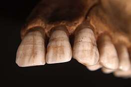
Enamel hypoplasia example
Linear enamel hypoplasia can be caused by a variety of factors, from genetic conditions to malnutrition and illnesses during childhood.
References
- May RL, Goodman AH, Meindl RS (September 1993). "Response of bone and enamel formation to nutritional supplementation and morbidity among malnourished Guatemalan children". American Journal of Physical Anthropology. 92 (1): 37–51. doi:10.1002/ajpa.1330920104. PMID 8238290.
- Miszkiewicz JJ (January 2015). "Linear Enamel Hypoplasia and Age‐at‐Death at Medieval (11th–16th Centuries) St. Gregory's Priory and Cemetery, Canterbury, UK". International Journal of Osteoarchaeology. 25 (1): 79–87. doi:10.1002/oa.2265.
- King T, Humphrey LT, Hillson S (November 2005). "Linear enamel hypoplasias as indicators of systemic physiological stress: evidence from two known age-at-death and sex populations from postmedieval London". American Journal of Physical Anthropology. 128 (3): 547–59. doi:10.1002/ajpa.20232. PMID 15861429.
- Berkovitz BK, Moxham BL (1978). Color Atlas and Textbook of Oral Anatomy, Histology and Embryology. London: Wolfe Medical Pub. pp. 79–88.
- Witzel C, Kierdorf U, Dobney K, Ervynck A, Vanpoucke S, Kierdorf H (July 2006). "Reconstructing impairment of secretory ameloblast function in porcine teeth by analysis of morphological alterations in dental enamel". Journal of Anatomy. 209 (1): 93–110. doi:10.1111/j.1469-7580.2006.00581.x. PMC 2100299. PMID 16822273.
- Guatelli-Steinberg D, Lukacs JR (1999). "Interpreting sex differences in enamel hypoplasia in human and non-human primates: Developmental, environmental, and cultural considerations". American Journal of Physical Anthropology. Suppl 29: 73–126. doi:10.1002/(SICI)1096-8644(1999)110:29+<73::AID-AJPA4>3.0.CO;2-K. PMID 10601984.
- Goodman AH, Rose JC (1990). "Assessment of systemic physiological perturbations from dental enamel hypoplasias and associated histological structures". American Journal of Physical Anthropology. 33: 59–110. doi:10.1002/ajpa.1330330506.
- King T, Hillson S, Humphrey LT (January 2002). "A detailed study of enamel hypoplasia in a post-medieval adolescent of known age and sex". Archives of Oral Biology. 47 (1): 29–39. doi:10.1016/s0003-9969(01)00091-7. PMID 11743929.
- Bartlett JD (September 2013). "Dental enamel development: proteinases and their enamel matrix substrates". ISRN Dentistry. 2013: 684607. doi:10.1155/2013/684607. PMC 3789414. PMID 24159389.
- Kanchan T, Machado M, Rao A, Krishan K, Garg AK (2015). "Enamel hypoplasia and its role in identification of individuals: A review of literature". Indian Journal of Dentistry. 6 (2): 99–102. doi:10.4103/0975-962X.155887. PMC 4455163. PMID 26097340.
- "What is Enamel Hypoplasia?".
- "A probable genetic origin for pitting enamel hypoplasia on the molars of Paranthropus robustus | Request PDF". ResearchGate. Retrieved 2019-03-28.
Further reading
- Pike-Tay A, Ma X, Hou Y, Liang F, Lin M, Peterson V (2016). "Combining Odontochronology, Tooth Wear Assessment, and Linear Enamel Hypoplasia (LEH) Recording to Assess Pig Domestication in Neolithic Henan, China". International Journal of Osteoarchaeology. 26 (1): 68–77. doi:10.1002/oa.2395.
- page 403 in Reitz EJ, Shackley M (2012). "Vertebrates". Environmental Archaeology. Manuals in Archaeological Method, Theory and Technique. pp. 383–422. doi:10.1007/978-1-4614-3339-2_12. ISBN 978-1-4614-3337-8.
This article is issued from Wikipedia. The text is licensed under Creative Commons - Attribution - Sharealike. Additional terms may apply for the media files.