Laser Doppler imaging
Laser Doppler imaging (LDI) can be used to measure superficial blood flow in the skin and other vascularized tissues. A laser Doppler perfusion imager functions by projecting a visible-to-infrared laser beam onto the surface of a tissue. When light interacts with moving blood cells a small portion of it is shifted in frequency according to the Doppler effect, detected, and converted into an electrical signal.[1]
| Laser Doppler imaging | |
|---|---|
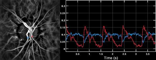
 Microangiography of the optic disc region of the human retina, by laser Doppler imaging. The image was rendered computationnally by optical wave propagation and measurement of optical fluctuations. | |
| Purpose | measure blood flow in eye |
| Based on | Digital holography |
By digital holography
The eye offers a unique opportunity for the non-invasive exploration of cardiovascular diseases. LDI by digital holography can measure blood flow in the retina and choroid.[2] In particular, the choroid is a highly vascularized tissue supplying the retinal pigment epithelium and photoreceptors. Yet investigating the anatomy and flow of the choroid remains challenging. LDI provides high-contrast visualization of local blood flow in choroidal vessels in humans, with a spatial resolution comparable to state-of-the-art indocyanine green angiography.[3] Differences in blood pressure drive the flow of blood throughout the circulation. The rate of mean blood flow depends on both blood pressure and the hemodynamic resistance to flow presented by the blood vessels. LDI can enable mapping of the local arterial resistivity index, and the possibility to perform unambiguous identification of retinal arteries and veins on the basis of their systole-diastole variations, and reveal ocular hemodynamics in human eyes.[4]
The local velocity of blood flow measured by laser Doppler holography in the digit (photoplethysmography) and the eye fundus has a pulse-shaped profile with time. These remote pulse wave measurements can be done clinically to reveal hemodynamics in arteries and veins and can be readily measured non-invasively.
Remote measurement of female sexual response
LDI provides a direct measure of female sexual response that does not require genital contact; signals are gathered at a depth of two to three millimetres below the skin's surface.[5] Two studies have suggested that LDI is a valid measure of female sexual arousal.[5][6] Waxman and Pukall[5] showed that LDI has discriminant validity; that is, it can differentiate sexual response from neutral, positive, and negative mood induced states. Compared to vaginal photoplethysmography (VPG), LDI is advantageous because it does not require genital manipulation or contact. Also, LDI provides a direct measure of vasocongestion and has an absolute unit of measurement, consisting of flux or units of blood flow. The disadvantages of LDI are that it cannot provide a continuous measure of sexual response and the laser Doppler perfusion imager is much more costly that other methods of genital sexual arousal assessment, such as VPG.[5]
See also
References
- Wardell, K., Jakobsson, A., & Nilsson, G. ["Laser Doppler perfusion imaging by dynamic light scattering"] "IEEE Transactions on Biomedical Engineering, 40, 309-316", 1993
- Puyo, L., M. Paques, M. Fink, J-A. Sahel, and M. Atlan. "In vivo laser Doppler holography of the human retina." Biomedical optics express 9, no. 9 (2018): 4113-4129.
- Puyo, Léo, Michel Paques, Mathias Fink, José-Alain Sahel, and Michael Atlan. "Choroidal vasculature imaging with laser Doppler holography." Biomedical optics express 10, no. 2 (2019): 995-1012.
- Puyo, Léo, Michel Paques, Mathias Fink, José-Alain Sahel, and Michael Atlan. "Waveform analysis of human retinal and choroidal blood flow with laser Doppler holography." Biomedical Optics Express 10, no. 10 (2019): 4942-4963.
- Waxman, S. E., & Pukall, C. F. ["Laser Doppler imaging of genital blood flow: A direct measure of female sexual arousal"], "The Journal of Sexual Medicine, 6(8), 2278-2285", 2009
- Stykes, S. J., MacLean, W. M. N., Sultana, S. R. ["Laser Doppler perfusion imaging: A method for measuring female sexual response"],"BJOG: International Journal of Obstetrics & Gynaecology, 113(5), 599–601", 2006
External links
| Wikimedia Commons has media related to Laser-doppler flowmetry. |