Jacalin-like lectin domain
In molecular biology, the jacalin-like lectin domain is a mannose-binding lectin domain with a beta-prism fold consisting of three 4-stranded beta-sheets, with an internal pseudo 3-fold symmetry. Some lectins in this group stimulate distinct T- and B-cell functions, such as Jacalin, which binds to the T-antigen and acts as an agglutinin. This domain is found in 1 to 6 copies in lectins. The domain is also found in the salt-stress induced protein from rice and an animal prostatic spermine-binding protein.
| Jacalin-like lectin domain | |||||||||
|---|---|---|---|---|---|---|---|---|---|
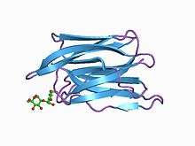 crystal structure of helianthus tuberosus lectin complexed to man(1-2)man | |||||||||
| Identifiers | |||||||||
| Symbol | Jacalin | ||||||||
| Pfam | PF01419 | ||||||||
| InterPro | IPR001229 | ||||||||
| SCOPe | 1jac / SUPFAM | ||||||||
| |||||||||
Database of jacalin like lectins and structure function relations.[1] Proteins containing this domain include:
- Jacalin, a tetrameric plant seed lectin and agglutinin from Artocarpus heterophyllus (jackfruit), which is specific for galactose.[2]
- Artocarpin, a tetrameric plant seed lectin from A. heterophyllus.[3]
- Lectin MPA, a tetrameric plant seed lectin and agglutinin from Maclura pomifera (Osage orange).[4]
- Heltuba lectin, a plant seed lectin and agglutinin from Helianthus tuberosus (Jerusalem artichoke).[5]
- Agglutinin from Calystegia sepium (Hedge bindweed).[6]
- Griffithsin, an anti-viral lectin from red algae (Griffithsia species).[7]
References
- Raval et al, "A database analysis of jacalin-like lectins: sequence–structure–function relationships" Glycobiology vol. 14 no. 12 pp. 1247–1263, 2004 http://glycob.oxfordjournals.org/content/14/12/1247.full.pdf
- Jeyaprakash AA, Geetha Rani P, Banuprakash Reddy G, Banumathi S, Betzel C, Sekar K, Surolia A, Vijayan M (August 2002). "Crystal structure of the jacalin-T-antigen complex and a comparative study of lectin-T-antigen complexes". J. Mol. Biol. 321 (4): 637–45. CiteSeerX 10.1.1.532.2424. doi:10.1016/S0022-2836(02)00674-5. PMID 12206779.
- Jeyaprakash AA, Srivastav A, Surolia A, Vijayan M (May 2004). "Structural basis for the carbohydrate specificities of artocarpin: variation in the length of a loop as a strategy for generating ligand specificity". J. Mol. Biol. 338 (4): 757–70. CiteSeerX 10.1.1.530.4331. doi:10.1016/j.jmb.2004.03.040. PMID 15099743.
- Lee X, Thompson A, Zhang Z, Ton-that H, Biesterfeldt J, Ogata C, Xu L, Johnston RA, Young NM (March 1998). "Structure of the complex of Maclura pomifera agglutinin and the T-antigen disaccharide, Galbeta1,3GalNAc". J. Biol. Chem. 273 (11): 6312–8. doi:10.1074/jbc.273.11.6312. PMID 9497359.
- Bourne Y, Zamboni V, Barre A, Peumans WJ, Van Damme EJ, Rouge P (December 1999). "Helianthus tuberosus lectin reveals a widespread scaffold for mannose-binding lectins". Structure. 7 (12): 1473–82. doi:10.1016/s0969-2126(00)88338-0. PMID 10647178.
- Bourne Y, Roig-Zamboni V, Barre A, Peumans WJ, Astoul CH, Van Damme EJ, Rouge P (January 2004). "The crystal structure of the Calystegia sepium agglutinin reveals a novel quaternary arrangement of lectin subunits with a beta-prism fold". J. Biol. Chem. 279 (1): 527–33. doi:10.1074/jbc.M308218200. PMID 14561768.
- Ziolkowska NE, O'Keefe BR, Mori T, Zhu C, Giomarelli B, Vojdani F, Palmer KE, McMahon JB, Wlodawer A (July 2006). "Domain-swapped structure of the potent antiviral protein griffithsin and its mode of carbohydrate binding". Structure. 14 (7): 1127–35. doi:10.1016/j.str.2006.05.017. PMC 7126681. PMID 16843894.
This article is issued from Wikipedia. The text is licensed under Creative Commons - Attribution - Sharealike. Additional terms may apply for the media files.