i-motif DNA
i-motif DNA, also called i-DNA, is a type of unusual structure of deoxyribonucleic acid, first found in the 1993 by Maurice Guéron and colleagues in École Polytechnique, France [1],and then discovered in cell nuclei in 2018, that is found in the nuclei of human cells. An abbreviation of intercalated motif, the four-strand i-motif is unlike the iconic double helix DNA.[2] I-motifs are four-stranded quadruplex structures formed by cytosine-rich DNA, similar to the G-quadruplex structures that guanine-rich DNA forms. C-rich DNA regions are common in gene regulation portions of the genome.[3] Recently, i-motifs were discovered in human cells and were shown to play a role in cell reproduction.[4] I-motifs have potential applications in nano-technology and nano-medicine, because size is more than 1 nm and less than 100 nm due to their unique pH sensitivity and have been used as biosensors, nanomachines, and molecular switches.[3]
History
The i-motif DNA was first found in the 1993 by Maurice Guéron and colleagues in École Polytechnique, France [1], but until 2018 had only ever been witnessed in vitro, not in living cells.[5] The latest discovery announced in 2018 was headed at the Garvan Institute of Medical Research and the University of New South Wales and funded by the National Health and Medical Research Council and the Australian Research Council. Co-leaders of the research team were A/Prof Daniel Christ and Professor Marcel Dinger.[2]
Discovery
Dr. Mahdi Zeraati at the Garvan Institute says the i-motif was discovered for its appearing and disappearing within the cell and distinctive connections via cytosines rather than base-pair letters. The research team developed a precise new tool to find the i-motif – a fragment of an antibody molecule – that could specifically recognise and attach to i-motifs "with a very high affinity".[2]
Structure
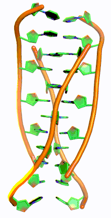
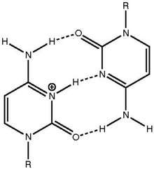
G-quadruplex DNA structures exist in G-rich areas of DNA. The complementary cytosine-rich DNA strand can also form a four-stranded quadruplex structure called i-motif. The cytosine base pairs in the i-motif structure form an intercalated and anti-parallel tetramer structure.[8] This structure is formed through non-Watson Crick base pairing C·C+ interactions of the cytosine base pairs.[8] C·C+ bonding is a type of Hoogsteen base pairing and actually has stronger base-pair interactions and base-pairing energy than the traditional Watson Crick G·C base pairing: 169.7 kJ/mol for C·C+ base pairing and 96.6 kJ/mol for G·C base-pairing.[8] The C·C+ bond has three hydrogen bonds: two between the hydrogen of the amines and the double bonded oxygens of the opposite cytosine, and one between the nitrogen and the hydrogen bound to the N+.[9] These base pairs cross-link two loops of DNA.[9] Given the hemi-protonated nature of the nitrogens in cytosine participating in C·C+ bonding, this base pairing traditionally is most stable in the range of pH=5–6, significantly lower than physiological pH (7.3).[10] However, recent work has found evidence of i-motif structures at near-neutral pH at room temperature.[11] Wright et al. discovered that the number of cytosines in the sequence determines if a C-rich sequence can form an i-motif structure at physiological pH, as does negative superhelicity[12] and molecular crowding.[11][13] At room temperature nucleotide tracts must be at least 5 cytosines long to fold into i-motifs at near-neutral pH; additional cytosines increase thermal stability.[11][14] Furthermore, intramolecular i-motif strands are most stable when there are at least 6 consecutive cytosine bases in the DNA strand while less than 6 consecutive cytosines in a strand favors intermolecular C·C+ pairings.[10] This pH-dependency of the i-motif structure is thought to be critical to its in vivo activity and function in the cell as pH is speculated to be the control mechanism used for i-motif formation and destruction.[14]
Biological function
I-motifs are postulated to play a role in gene regulation and expression in the cell.[15] Large tracts of G/C-rich DNA exist near transcriptional start sites in the genome, and are present in a wide variety of organismal genomes, suggesting that C-rich tracts have a biological function.[16] Furthermore, many proteins and ligands fundamental to gene expression recognize C-rich oligonucleotides, such as Poly-C-binding protein (PCBP) and heterogeneous nuclear ribonucleoprotein K (HNRPK).[16] Until recently it was not clear whether I-motifs naturally existed in DNA due to their low stability at physiological pH;[16] i-motifs were discovered in vivo in human DNA in 2018.[17] Human telomeric DNA (hTelo) can form an i-motif secondary structure in vitro.[18] Zeraati et al. confirmed the presence of i-motif hTelo in human DNA using a fluorescent marker called iMab.[17] Furthermore, i-motif hTelo was found within regulatory regions of the genome during the late G1 phase of the cell reproduction cycle, indicating that i-motifs are involved in gene promotion and regulation.[17] I-motifs may also act as molecular scaffolds to aid in transcription factor binding during gene transcription by aiding promoters such as BCL2 in attaching to the correct DNA sequence.[17][19] Increased i-motif expression did not coincide with increased G-quadruplex expression, which instead increases during the S phase.[17] This indicates that G-quadruplex and i-motifs are not complementary structures despite their complementary sequences and are instead mutually exclusive and serve opposite roles in gene expression regulation.[17]
Applications
I-motifs are particularly useful due to their unique sensitivity to changes in acidity near physiological pH. A study at the University of Bonn used I-motif folding to tighten and loosen a ring of DNA. A circular ring of DNA was synthesized, with certain regions of C-rich DNA.[20] At a pH of 5, these regions contracted to form i-motifs, tightening the ring in a fashion similar to closing a trash bag. At a pH of 8 the I-motif regions collapsed back into their linear forms, relaxing the ring. DNA rings that can tighten and loosen based on pH can be used to build more complex structures of interlocking DNA like catenanes and rotaxanes.[20] These DNA structures can function as molecular switches. Another study showed that single-walled carbon nanotubes (SWNTs), commonly used to carry drugs in the body, induce I-motif formation in human telomeric DNA.[21] Researches modified C-rich human telomeric DNA by attaching a redox active methylene blue group to the 3′ end and an electrode to the 5′ end. In the I-motif conformation this modified DNA strand produces a large increase in Faradaic current. This biosensor only reacts to SWNTs, allowing researchers to detect a specific type of carbon nanotube with a direct detection limit of 0.2 ppm.[21]
See also
References
- Gehring, Kalle; Leroy, Jean-Louis; Guéron, Maurice. "A tetrameric DNA structure with protonated cytosine-cytosine base pairs". Nature. 363: 561–565. doi:10.1038/363561a0.
- Public statement: "Found: A new form of DNA in our cells."
- Benabou, S.; Aviñó, A.; Eritja, R.; González, C.; Gargallo, R. (2014). "Fundamental aspects of the nucleic acid i-motif structures" (PDF). RSC Adv. 4 (51): 26956–26980. doi:10.1039/c4ra02129k. ISSN 2046-2069.
- Zeraati, Mahdi; Langley, David B.; Schofield, Peter; Moye, Aaron L.; Rouet, Romain; Hughes, William E.; Bryan, Tracy M.; Dinger, Marcel E.; Christ, Daniel (23 April 2018). "I-motif DNA structures are formed in the nuclei of human cells". Nature Chemistry. 10 (6): 631–637. doi:10.1038/s41557-018-0046-3. ISSN 1755-4330. PMID 29686376.
- "Scientists Have Confirmed a New DNA Structure Inside Human Cells", article by Peter Dockrill 23 April 2018
- Snoussi, K.; Nonin-Lecomte, S.; Lerou, J.L. (30 May 2001). "THE RNA I-MOTIF". doi:10.2210/pdb1i9k/pdb. Cite journal requires
|journal=(help) - Gurung, Sarah P.; Schwarz, Christine; Hall, James P.; Cardin, Christine J.; Brazier, John A. (2015). "The importance of loop length on the stability of i-motif structures". Chemical Communications. 51 (26): 5630–5632. doi:10.1039/c4cc07279k. ISSN 1359-7345. PMC 4384421. PMID 25686374.
- Benabou, S.; Aviñó, A.; Eritja, R.; González, C.; Gargallo, R. (2014). "Fundamental aspects of the nucleic acid i-motif structures" (PDF). RSC Adv. 4 (51): 26956–26980. doi:10.1039/c4ra02129k. ISSN 2046-2069.
- Zhang, Xi Yuan; Luo, Hong Qun; Li, Nian Bing (15 June 2014). "Crystal violet as an i-motif structure probe for reversible and label-free pH-driven electrochemical switch". Analytical Biochemistry. 455: 55–59. doi:10.1016/j.ab.2014.03.015. ISSN 0003-2697. PMID 24699211.
- Li, Tao; Famulok, Michael (22 January 2013). "I-Motif-Programmed Functionalization of DNA Nanocircles". Journal of the American Chemical Society. 135 (4): 1593–1599. doi:10.1021/ja3118224. ISSN 0002-7863. PMID 23312021.
- Wright, Elisé P.; Huppert, Julian L.; Waller, Zoë A. E. (9 February 2017). "Identification of multiple genomic DNA sequences which form i-motif structures at neutral pH". Nucleic Acids Research. 45 (6): 2951–2959. doi:10.1093/nar/gkx090. ISSN 0305-1048. PMC 5605235. PMID 28180276.
- Sun, Daekyu; Hurley, Laurence H. (14 May 2009). "The Importance of Negative Superhelicity in Inducing the Formation of G-Quadruplex and i-Motif Structures in the c-Myc Promoter: Implications for Drug Targeting and Control of Gene Expression". Journal of Medicinal Chemistry. 52 (9): 2863–2874. doi:10.1021/jm900055s. ISSN 0022-2623. PMC 2757002. PMID 19385599.
- Cui, Jingjing; Waltman, Phillip; Le, Vu; Lewis, Edwin (15 October 2013). "The Effect of Molecular Crowding on the Stability of Human c-MYC Promoter Sequence I-Motif at Neutral pH". Molecules. 18 (10): 12751–12767. doi:10.3390/molecules181012751. ISSN 1420-3049. PMC 6270392. PMID 24132198.
- Zeraati, Mahdi; Langley, David B.; Schofield, Peter; Moye, Aaron L.; Rouet, Romain; Hughes, William E.; Bryan, Tracy M.; Dinger, Marcel E.; Christ, Daniel (23 April 2018). "I-motif DNA structures are formed in the nuclei of human cells". Nature Chemistry. 10 (6): 631–637. doi:10.1038/s41557-018-0046-3. ISSN 1755-4330. PMID 29686376.
- Wright, Elisé P.; Huppert, Julian L.; Waller, Zoë A. E. (9 February 2017). "Identification of multiple genomic DNA sequences which form i-motif structures at neutral pH". Nucleic Acids Research. 45 (6): 2951–2959. doi:10.1093/nar/gkx090. ISSN 0305-1048. PMC 5605235. PMID 28180276.
- Benabou, S.; Aviñó, A.; Eritja, R.; González, C.; Gargallo, R. (2014). "Fundamental aspects of the nucleic acid i-motif structures" (PDF). RSC Adv. 4 (51): 26956–26980. doi:10.1039/c4ra02129k. ISSN 2046-2069.
- Zeraati, Mahdi; Langley, David B.; Schofield, Peter; Moye, Aaron L.; Rouet, Romain; Hughes, William E.; Bryan, Tracy M.; Dinger, Marcel E.; Christ, Daniel (23 April 2018). "I-motif DNA structures are formed in the nuclei of human cells". Nature Chemistry. 10 (6): 631–637. doi:10.1038/s41557-018-0046-3. ISSN 1755-4330. PMID 29686376.
- Manzini, G.; Yathindra, N.; Xodo, L. E. (11 November 1994). "Evidence for intramolecularly folded i-DNA structures in biologically relevant CCC-repeat sequences". Nucleic Acids Research. 22 (22): 4634–4640. doi:10.1093/nar/22.22.4634. ISSN 0305-1048. PMC 308511. PMID 7984411.
- Kang, Hyun-Jin; Kendrick, Samantha; Hecht, Sidney M.; Hurley, Laurence H. (7 March 2014). "The Transcriptional Complex Between the BCL2 i-Motif and hnRNP LL Is a Molecular Switch for Control of Gene Expression That Can Be Modulated by Small Molecules". Journal of the American Chemical Society. 136 (11): 4172–4185. doi:10.1021/ja4109352. ISSN 0002-7863. PMC 3985447. PMID 24559432.
- Li, Tao; Famulok, Michael (22 January 2013). "I-Motif-Programmed Functionalization of DNA Nanocircles". Journal of the American Chemical Society. 135 (4): 1593–1599. doi:10.1021/ja3118224. ISSN 0002-7863. PMID 23312021.
- Peng, Yinghua; Wang, Xiaohui; Xiao, Yi; Feng, Lingyan; Zhao, Chao; Ren, Jinsong; Qu, Xiaogang (30 September 2009). "i-Motif Quadruplex DNA-Based Biosensor for Distinguishing Single- and Multiwalled Carbon Nanotubes". Journal of the American Chemical Society. 131 (38): 13813–13818. doi:10.1021/ja9051763. ISSN 0002-7863. PMID 19736925.