Ground-glass opacity
In radiology, ground glass opacity (GGO) is a nonspecific finding on radiographs and computed tomography (CT) scans. It consists of a hazy opacity that does not obscure the underlying bronchial structures or pulmonary vessels, and that indicates a partial filling of air spaces in the lungs by exudate or transudate, as well as interstitial thickening or partial collapse of lung alveoli.[1]:95

Differential diagnosis
The differential diagnosis of the many causes of GGO includes pulmonary edema, infections (including severe acute respiratory syndrome coronavirus 2 (COVID-19),[2] cytomegalovirus and Pneumocystis jirovecii pneumonia), various noninfectious interstitial lung diseases (such as hypersensitivity pneumonitis, Hamman-Rich syndrome), diffuse alveolar hemorrhage, cryptogenic organizing pneumonia, and pulmonary contusion.[3]
Reversed halo sign
A reversed halo sign is a central ground-glass opacity surrounded by denser consolidation. Criteria include that the consolidation should form more than three-fourths of a circle and be at least 2 mm thick.[4] It is suggestive of cryptogenic organizing pneumonia,[5] but is only seen in about 20% of individuals with this condition.[4] It can also be present in lung infarction where the halo consists of hemorrhage,[6] as well as in infectious diseases such as paracoccidioidomycosis, tuberculosis, zygomycosis, and aspergillosis, as well as in granulomatosis with polyangiitis, lymphomatoid granulomatosis, and sarcoidosis.[7]
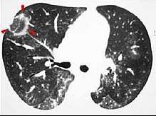 CT scan with reversed halo sign in a case of cryptogenic organizing pneumonia.
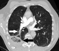 CT scan of a reverse halo sign (black arrow) due to lung infarction because of chronic pulmonary embolism (white arrow).
References
- Hodler, J.; von Schulthess, G. K.; Zollikofer, C. L. (2007). Diseases of the Heart, Chest & Breast: Diagnostic Imaging and Interventional Techniques. Berlin/Heidelberg: Springer. p. 95. ISBN 9788847006331.
- Bernheim A, Mei X, Huang M, Yang Y, Fayad ZA, Zhang N, et al. (February 2020). "Chest CT Findings in Coronavirus Disease-19 (COVID-19): Relationship to Duration of Infection". Radiology: 200463. doi:10.1148/radiol.2020200463. PMID 32077789.
- Jannette Collins, MD; Eric J. Stern, MD (1998). "Ground glass opacity on CT scanning of the chest: What does it mean?" (PDF). Applied Radiology. Archived from the original (PDF) on 18 May 2012. Retrieved 1 February 2012.
- Radswiki; et al. "Reversed halo sign (lungs)". Radiopaedia. Retrieved 2 January 2018.
- Brett M. Elicker, W. Richard Webb (2012). Fundamentals of High-Resolution Lung CT: Common Findings, Common Patterns, Common Diseases, and Differential Diagnosis. Lippincott Williams & Wilkins. ISBN 9781469824796.
- Wu, George; Schmit, Berndt; Arteaga, Veronica; Palacio, Diana (2017). "Medical image of the week: pulmonary infarction- the "reverse halo sign"". Southwest Journal of Pulmonary and Critical Care. 15 (4): 162–163. doi:10.13175/swjpcc124-17. ISSN 2160-6773.
- D Karthikeyan (2013). High Resolution Computed Tomography of the Lungs: A Practical Guide. JP Medical Ltd. p. 256. ISBN 9789350904084.