Denis Baylor
Denis Aristide Baylor (born January 30, 1940) is a Professor of Neurobiology at Stanford University. He is best known for his research on retinal cells and developing a technique to study electrical activity within cells.[2] This technique has become commonly used today by many scientists.[2] Baylor has received numerous awards for his work including an appointment as a foreign member of the Royal Society of London, being appointed as a member of the National Academy of Sciences, becoming a fellow of American Academy of Arts and Sciences, and receiving the MERIT Award from the National Eye Institute.[1]
Denis Baylor | |
|---|---|
| Nationality | U.S. |
| Citizenship | American |
| Alma mater |
|
| Awards |
|
| Scientific career | |
| Fields | Neurobiology Cell Biology [1] |
| Institutions |
|
| Doctoral advisor | John Nicholls |
Early life and education
Denis Baylor was born on January 30, 1940 in Oskaloosa, Iowa.[3] Baylor received his BA in Chemistry from Knox College in 1961, where he graduated Magna Cum Laude.[1] During his undergraduate career he was a member of Phi Beta Kappa and Alpha Omega Alpha Honor Societies. In 1965, Baylor received his M.D. from Yale School of Medicine, where he graduated Cum Laude.[4] He continued his education by completing a Postdoctoral Fellow in Physiology with John Nicholls between 1965 and 1968.[1]
Research and career
Academic posts
After completing his Postdoctoral Fellow, Baylor worked as a lab associate at the National Institute of Neurological Disorders and Stroke from 1968 to 1970. Baylor was an Associate professor of Physiology at the University of Colorado Medical School.[1] He then moved to Stanford University where he was an Associate Professor of Physiology up to 1975.[1]. Baylor was then an Associate Professor of Neurobiology between 1975 and 1978. In 1978, he became a Professor of Neurobiology at Stanford until 2001, when he became a Professor Emeritus of Neurobiology at Stanford.[1] Throughout his career, Baylor has served on the visiting committee at Harvard Medical School, the editorial board for The Journal of Physiology, Neuron, Journal of Neurophysiology, Visual Neuroscience, and The Journal of Neuroscience. He has also been on multiple medical and scientific advisory boards, along with being a Trustee for multiple foundations.[1][5]
Research interests and selected publications
One of Baylor's first large works was published in 1985 by the National Academy of Science in an article titled “Interaction of Hydrolysis-Resistant Analogs of Cyclic
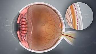
GMP with the Phosphodiesterase and Light-Sensitive Channel of Retinal Rod Outer Segments.” Throughout this study, a series of nonhydrolyzable versions of cGMP were used to test the light sensitive gating mechanism of the rods.[6] The hydrolysis of the cGMP allows for the normal light response of the rods, opening channels for the entrance of light and closing in its absence. The study monitored the electrodes that were flowing throughout the cell with different nonhydrolyzable analogs. It was found that when the channel receptor was bound to the homologue of cGMP there was an increase in the dark current and a much slower overall response to light.[6] The study also found that whether a cGMP or its homologue was used, an increased concentration present could cause the rods to have slower responses as there was less control on the ability to open the gate quickly. Overall, this study provided insight into how light travels to the rods, and is used and processed by the rods within the eye.[6]
In 1991, Baylor published “Synchronous bursts of action potentials in ganglion cells of the developing mammalian retina” where he studied the visual systems of mammals.[7] The study used stimulus recordings to measure the action potential of the optic nerve fiber, and measured electrical movement of the neurons into the different eye specific layers of the retina[7]. The recorded electrical activity from the neurons excited many ganglion cells. This study illustrated that the fired neurons created the connection between the retina and the geniculate nucleus. Overall, this study was a breakthrough in proving how light can move through the eye and be received by the brain so that images and the environment can be perceived rapidly.[7]
Baylor's study on “Multi-neuronal signals from the retina: acquisition and analysis” utilized a new method to observe the many electrical activities of all the neurons in a section of the retina of the eye.[8] This study focused on how the eye processes information it receives despite having so many overlapping nerve fibers and networks. The extracellular action potentials are measured giving an idea as to the arrangement of the ganglion cells, which was followed by the measure of the voltage of the signals.[8] In addition, this study utilized visual methods to characterize a neuron's response to stimuli. The results from these two methods were combined and correlations were created between the electrical activity and visual responses to determine the different functions of the ganglion cells when prompted with a stimulus. Overall the new methods used in this study could help future studies publish detailed information about how optic nerve fibers work.[8]
In 1996, Baylor's article “How photons start vision” was published in the Proceedings of the National Academy of Sciences of the United States of America. This paper addressed the process of shutting down the transduction pathway that occurs in the response to light stimuli.[9] When a photon enters the eye, it is sent to the back of the eye and is absorbed by rods and cones. These cells are excited by the photon and as a response hydrolyze the GTP and cGMP. This finding proved to be the rate limiting step.[9] This causes gated channels to close and create a polarity. The signal can then be sent through the nerve cells to the brain where the image is processed. The study utilized lab controlled cells that could easily show the presence and concentration of nucleotides and calcium.[9] It showed that to end this signal amplification there has to be a drop in Ca2+ concentrations, which results in a negative feedback to return the cell back to its original state with GTP and cGMP, unhydrolyzed.[9]
His more recent publications focus on genetically targeting cells and observing what happens to their function with a deletion, over-expression, or insertion.[9] This information can give more detail into the job of individual cells, and their roles in processing images.
Awards and honors
Baylor has received many awards and honors throughout his career in neurobiology and cellular biology. He was awarded the Sinsheimer Foundation Award for Medical Research in 1975. He received the Mathilde Solowey Award in NeuroSciences in 1978.[1] He also received the Proctor medal from the Association for Research in Vision and Ophthalmology in 1986. In 1988, Dr. Baylor received the Kayser International Award from the Retina Research Foundation. Along with these awards, he has received appointments to the Royal Society of London (2003), the National Academy of Science (1993), and became a fellow of American Academy of Arts and Sciences (1992).[1][2][4]
References
- "Denis Baylor's Profile | Stanford Profiles". profiles.stanford.edu. Retrieved 2020-03-31.
- "Search | Royal Society". royalsociety.org. Retrieved 2020-03-31.
- Baylor, Denis "Curriculum Vitae" via https://profiles.stanford.edu/denis-baylor
- "Denis Baylor". www.nasonline.org. Retrieved 2020-03-31.
- "1996 | The Grass Foundation". www.grassfoundation.org. Retrieved 2020-03-31.
- Zimmerman, Anita L.; Yamanaka, Gregory; Eckstein, Fritz; Baylor, Denis A.; Stryer, Lubert (1985). "Interaction of Hydrolysis-Resistant Analogs of Cyclic GMP with the Phosphodiesterase and Light-Sensitive Channel of Retinal Rod Outer Segments". Proceedings of the National Academy of Sciences of the United States of America. 82 (24): 8813–8817. Bibcode:1985PNAS...82.8813Z. doi:10.1073/pnas.82.24.8813. ISSN 0027-8424. JSTOR 26683. PMC 391528. PMID 2417228.
- Meister, M; Wong, R.; Baylor, D.; Shatz, C. (1991-05-17). "Synchronous bursts of action potentials in ganglion cells of the developing mammalian retina". Science. 252 (5008): 939–943. Bibcode:1991Sci...252..939M. doi:10.1126/science.2035024. ISSN 0036-8075. PMID 2035024.
- Meister, Markus; Pine, Jerome; Baylor, Denis A. (1994). "Multi-neuronal signals from the retina: acquisition and analysis". Journal of Neuroscience Methods. 51 (1): 95–106. doi:10.1016/0165-0270(94)90030-2. PMID 8189755.
- Burns, Marie E; Baylor, Denis A (2001). "Activation, Deactivation, and Adaptation in Vertebrate Photoreceptor Cells". Annual Review of Neuroscience. 24 (1): 779–805. doi:10.1146/annurev.neuro.24.1.779. ISSN 0147-006X. PMID 11520918.