C11orf54
Chromosome 11 open reading frame 54 (C11orf54) is a protein that in humans is encoded by the C11orf54 gene.[5] The "Homo sapiens" gene, C11orf54 is also known as PTD012 and PTOD12. C11orf54 exhibits hydrolase activity on p-nitrophenyl acetate and acts on ester bonds, though the overall function is still not fully understood by the scientific community. The protein is highly conserved with the most distant homolog found is in bacteria.[6]
Gene
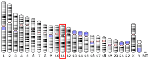
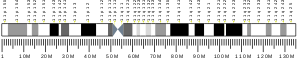
C11orf54 is located on chromosome 11 at 11q21. Common aliases of the gene are PTD012 and PT0D12. The gene consists 13 exons and spans 23730 bp. C11orf54 is flanked by TAF1D and MED17.[6]
mRNA
The protein ester hydrolase c11orf54 exists as a monomer and is composed of 315 amino acids. There are 6 isoforms for C11orf54. See table 1.[6]
| Variant | Isoform | Length (bp) | Accession Number |
|---|---|---|---|
| 1 | ester hydrolase C11orf54 isoform a | 2726 | NM_001286067.1 |
| 2 | ester hydrolase C11orf54 isoform a | 2589 | NM_001286068.1 |
| 3 | ester hydrolase C11orf54 isoform a | 2594 | NM_001286069.1 |
| 4 | ester hydrolase C11orf54 isoform b | 2444 | NM_014039.3 |
| 5 | ester hydrolase C11orf54 isoform c | 2442 | NM_001286070.1 |
| 6 | ester hydrolase C11orf54 isoform d | 2417 | NM_001286071.1 |
The amino acid sequence contains the domain of unknown function 1907. Found in this transcript is the HxHxxxxxxxxxH motif which coordinates the zinc ion involved in the hydrolase activity.[7] An LR nest motif is found at lys262 and Arg263. The LR nest motif forms hydrogen bonds between the NH groups and anions; an acetate anion is coordinated with the LR nest.[8]
Protein
Primary sequence
Table 2 shows the different characteristics of the protein sequence throughout humans and other orthologs.[9]
| Organism | Molecular Weight (kiloDalton) | Isoelectric point | High Bias Amino Acids | Repeats |
|---|---|---|---|---|
| Human | 35.1 | 5.9 | F | AEFS |
| Mouse | 35.0 | 5.9 | H | None |
| 13 Lined Ground Squirrel | 35.1 | 6.0 | F,H | PAEF |
| Giant Panda | 35.2 | 6.5 | F | PAEF |
Secondary structure
The protein of C11orf54 exists as a monomer in solution. The protein assumes a globular shape of 20 beta strands and 4 alpha helices, containing 9 antiparallel beta strands forming a beta screw region. The β-screw region of C11orf54 has structural similarity to the cyclic adenosine 3′,5′-monophosphate (cAMP) binding domain of the regulatory subunit of protein kinase A. A zinc ion is bound to the HxHxxxxxxxxxH motif found in the sequence.[7]
Subcellular localization
C11orf54 is predicted to be localized 60.9% in the cytoplasm, 21.7% in the nucleus, 13.0% mitochondrial and 4.3% in the Golgi Apparatus.[10]
Homology
Paralogs
There are no paralogs for C11orf54.[5]
Orthologs
The protein Ester Hydrolase C11orf54 has many orthologs (see table.) It is highly conserved (60-100% identity) in mammals, reptiles, birds, and fish. The protein is moderately conserved (30-59.99% identity) in invertebrates, amphibia, Cnidaria, Mollusca, fungi and bacteria. It is not conserved in archaea.[9] The most distant orthologs are bacteria. Figure 2 shows the unrooted phylogenetic tree of a few of C11orf54’s orthologs.[5]
| Species | Common Name | Class | Accession Number | Percent Identity | Divergence (MYA median) |
|---|---|---|---|---|---|
| Microtus ochrogaster | Prairie Vole | mammalia | XP_005346877.1 | 87.0 | 88 |
| Chelonia mydas | Green Sea Turtle | reptilia | XP_007069537.1 | 72.8 | 320 |
| Xenopus tropicalis | Burmese Python | reptilia | XP_007434894.1 | 70.9 | 320 |
| Python bivittatus | Red Junglefowl | Ave | NP_001264206.1 | 73.4 | 320 |
| Gallus gallus | Common Cuckoo | Ave | XP_009564677.1 | 72.5 | 320 |
| Cuculus canorus | Southern Platyfish | Actinopterygii | XP_005800827.1 | 65.2 | 432 |
| Xiphophorus maculatus | Zebrafish | Actinopterygii | NP_997781.1 | 62.4 | 432 |
| Danio rerio | Acorn Worm | Enteropneusta | XP_002738479.1 | 55.6 | 627 |
| Saccoglossus kowalevskii | Atlantic Horseshoe Crab | Merostomata | XP_013785734.1 | 56.6 | 758 |
| Limulus polyphemus | Western Clawed Frog | Amphibia | XP_012812415.1 | 55.1 | 353 |
| Crassostrea gigas | Pacific Oyster | Bivalvia | XP_011412414.1 | 50.0 | 758 |
| Tribolium castaneum | Red Flour Beetle | Insecta | XP_968861.1 | 49.0 | 758 |
| Drosophila bipectinata | Fruitfly | Insecta | XP_017103988.1 | 46.0 | 758 |
| Megachile rotundata | Alfalfa leafcutter bee | Insecta | XP_003702672.1 | 44.8 | 758 |
| Zymoseptoria brevis | fungi | Dothideomycetes | KJX93246.1 | 36.5 | 1150 |
| Cladophialophora carrionii | fungi | Dothideomycetes | OCT48531.1 | 35.8 | 1150 |
| Alternaria alternata | fungi | Dothideomycetes | XP_018384285.1 | 36.2 | 1150 |
| Candidatus Pelagibacter ubique | bacteria | Bacteria | WP_075504325.1 | 34.5 | 4090 |
| Pelagibacteraceae bacterium | bacteria | Bacteria | OCW82973.1 | 34.1 | 4090 |
Function
C11orf54's coordination with a zinc ion through three histidines and an acetate anion is likely to point to a function of the protein being an enzymatic reaction as an ester hydrolase. The protein has a high turnover number when reacted with p-nitrophenyl acetate (0.042 sec−1) as compared to a 1 sec−1 turnover rate found in another enzyme (bovine carbonic anhydrase II) that reacts with p-nitrophenyl acetate.[7]
Interacting Proteins
| Protein Name | Abbreviation |
|---|---|
| Ubiquitin C | UBC |
| Collagen, type IV, alpha 3 | COL4A3 |
| Thyroid Hormone Receptor Interactor 13 | TRIP13 |
| DEAD (Asp-Glu-Ala-Asp) box polypeptide 60-like | DDX60L |
| Glutamine-fructose-6-phosphate transaminase 2 | GFPT2 |
| Superkiller viralicidic activity 2-like (S. cerevisiae) | SKIV2L |
| OTU domain, ubiquitin aldehyde binding 1 | OTUB1 |
References
- GRCh38: Ensembl release 89: ENSG00000182919 - Ensembl, May 2017
- GRCm38: Ensembl release 89: ENSMUSG00000031938 - Ensembl, May 2017
- "Human PubMed Reference:". National Center for Biotechnology Information, U.S. National Library of Medicine.
- "Mouse PubMed Reference:". National Center for Biotechnology Information, U.S. National Library of Medicine.
- "Entrez Gene: C11orf54 chromosome 11 open reading frame 54".
- "C11orf54". NCBI Gene. NCBI (National Center for Biotechnology Information).
- Manjasetty BA, Büssow K, Fieber-Erdmann M, Roske Y, Gobom J, Scheich C, Götz F, Niesen FH, Heinemann U (April 2006). "Crystal structure of Homo sapiens PTD012 reveals a zinc-containing hydrolase fold". Protein Science. 15 (4): 914–20. doi:10.1110/ps.052037006. PMC 2242484. PMID 16522806.
- Langton MJ, Serpell CJ, Beer PD (2016). "Anion Recognition in Water: Recent Advances from a Supramolecular and Macromolecular Perspective". Angewandte Chemie International Edition. 55 (6): 1974–87. doi:10.1002/anie.201506589. PMC 4755225. PMID 26612067.
- Subramaniam S (1998). "The Biology Workbench--a seamless database and analysis environment for the biologist". Proteins. 32 (1): 1–2. doi:10.1002/(SICI)1097-0134(19980701)32:1<1::AID-PROT1>3.0.CO;2-Q. PMID 9672036.
- Briesemeister S, Rahnenführer J, Kohlbacher O (2010). "Going from where to why–interpretable prediction of protein subcellular localization". Bioinformatics. 26 (9): 1232–8. doi:10.1093/bioinformatics/btq115. PMC 2859129. PMID 20299325.
- Blom N, Gammeltoft S, Brunak S (1999). "Sequence and structure-based prediction of eukaryotic protein phosphorylation sites". Journal of Molecular Biology. 294 (5): 1351–62. doi:10.1006/jmbi.1999.3310. PMID 10600390.
- Gupta R, Brunak S (2002). "Prediction of glycosylation across the human proteome and the correlation to protein function". Pacific Symposium on Biocomputing. Pacific Symposium on Biocomputing: 310–22. doi:10.1142/9789812799623_0029. ISBN 978-981-02-4777-5. PMID 11928486.
- Uhlén M, Fagerberg L, Hallström BM, Lindskog C, Oksvold P, Mardinoglu A, et al. (January 2015). "Proteomics. Tissue-based map of the human proteome". Science. 347 (6220): 1260419. doi:10.1126/science.1260419. PMID 25613900.
- Franceschini A, Szklarczyk D, Frankild S, Kuhn M, Simonovic M, Roth A, Lin J, Minguez P, Bork P, von Mering C, Jensen LJ (2013). "STRING v9.1: protein-protein interaction networks, with increased coverage and integration". Nucleic Acids Research. 41 (Database issue): D808–15. doi:10.1093/nar/gks1094. PMC 3531103. PMID 23203871.
Further reading
- Maruyama K, Sugano S (January 1994). "Oligo-capping: a simple method to replace the cap structure of eukaryotic mRNAs with oligoribonucleotides". Gene. 138 (1–2): 171–4. doi:10.1016/0378-1119(94)90802-8. PMID 8125298.
- Suzuki Y, Yoshitomo-Nakagawa K, Maruyama K, Suyama A, Sugano S (October 1997). "Construction and characterization of a full length-enriched and a 5'-end-enriched cDNA library". Gene. 200 (1–2): 149–56. doi:10.1016/S0378-1119(97)00411-3. PMID 9373149.
- Wiemann S, Weil B, Wellenreuther R, Gassenhuber J, Glassl S, Ansorge W, Böcher M, Blöcker H, Bauersachs S, Blum H, Lauber J, Düsterhöft A, Beyer A, Köhrer K, Strack N, Mewes HW, Ottenwälder B, Obermaier B, Tampe J, Heubner D, Wambutt R, Korn B, Klein M, Poustka A (March 2001). "Toward a catalog of human genes and proteins: sequencing and analysis of 500 novel complete protein coding human cDNAs". Genome Research. 11 (3): 422–35. doi:10.1101/gr.GR1547R. PMC 311072. PMID 11230166.
- Simpson JC, Wellenreuther R, Poustka A, Pepperkok R, Wiemann S (September 2000). "Systematic subcellular localization of novel proteins identified by large-scale cDNA sequencing". EMBO Reports. 1 (3): 287–92. doi:10.1093/embo-reports/kvd058. PMC 1083732. PMID 11256614.
- Wan D, Gong Y, Qin W, Zhang P, Li J, Wei L, Zhou X, Li H, Qiu X, Zhong F, He L, Yu J, Yao G, Jiang H, Qian L, Yu Y, Shu H, Chen X, Xu H, Guo M, Pan Z, Chen Y, Ge C, Yang S, Gu J (November 2004). "Large-scale cDNA transfection screening for genes related to cancer development and progression". Proceedings of the National Academy of Sciences of the United States of America. 101 (44): 15724–9. Bibcode:2004PNAS..10115724W. doi:10.1073/pnas.0404089101. PMC 524842. PMID 15498874.
- Rual JF, Venkatesan K, Hao T, Hirozane-Kishikawa T, Dricot A, Li N, Berriz GF, Gibbons FD, Dreze M, Ayivi-Guedehoussou N, Klitgord N, Simon C, Boxem M, Milstein S, Rosenberg J, Goldberg DS, Zhang LV, Wong SL, Franklin G, Li S, Albala JS, Lim J, Fraughton C, Llamosas E, Cevik S, Bex C, Lamesch P, Sikorski RS, Vandenhaute J, Zoghbi HY, Smolyar A, Bosak S, Sequerra R, Doucette-Stamm L, Cusick ME, Hill DE, Roth FP, Vidal M (October 2005). "Towards a proteome-scale map of the human protein-protein interaction network". Nature. 437 (7062): 1173–8. Bibcode:2005Natur.437.1173R. doi:10.1038/nature04209. PMID 16189514.