ATP synthase subunit C
ATPase, subunit C of Fo/Vo complex is the main transmembrane subunit of V-type, A-type and F-type ATP synthases.
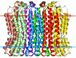 V-type sodium ATPase from Enterococcus hirae. Calculated hydrocarbon boundaries of the lipid bilayer are shown by red and blue dots | |||||||||
| Identifiers | |||||||||
|---|---|---|---|---|---|---|---|---|---|
| Symbol | ATP-synt_C | ||||||||
| Pfam | PF00137 | ||||||||
| InterPro | IPR002379 | ||||||||
| PROSITE | PDOC00526 | ||||||||
| SCOPe | 1aty / SUPFAM | ||||||||
| OPM superfamily | 5 | ||||||||
| OPM protein | 2bl2 | ||||||||
| |||||||||
ATPases (or ATP synthases) are membrane-bound enzyme complexes/ion transporters that combine ATP synthesis and/or hydrolysis with the transport of protons across a membrane. ATPases can harness the energy from a proton gradient, using the flux of ions across the membrane via the ATPase proton channel to drive the synthesis of ATP. Some ATPases work in reverse, using the energy from the hydrolysis of ATP to create a proton gradient. There are different types of ATPases, which can differ in function (ATP synthesis and/or hydrolysis), structure (F-, V- and A-ATPases contain rotary motors) and in the type of ions they transport.[1][2]
- F-ATPases (F1Fo ATPases) in mitochondria, chloroplasts and bacterial plasma membranes are the prime producers of ATP, using the proton gradient generated by oxidative phosphorylation (mitochondria) or photosynthesis (chloroplasts).
- V-ATPase (V1Vo ATPases) are primarily found in eukaryotic vacuoles, catalysing ATP hydrolysis to transport solutes and lower pH in organelles.
- A-ATPases (A1Ao ATPases) are found in Archaea and function like F-ATPases.
- P-ATPases (E1E2 ATPases) are found in bacteria and in eukaryotic plasma membranes and organelles, and function to transport a variety of different ions across membranes.
- E-ATPases are cell-surface enzymes that hydrolyse a range of NTPs, including extracellular ATP.
The F-ATPases (or F1Fo ATPases) and V-ATPases (or V1Vo ATPases) are each composed of two linked complexes: the F1 or V1 complex contains the catalytic core that synthesizes/hydrolyses ATP, and the Fo or Vo complex that forms the membrane-spanning pore. The F- and V-ATPases all contain rotary motors, one that drives proton translocation across the membrane and one that drives ATP synthesis/hydrolysis.[3][4]
Subunit C (also called subunit 9, or proteolipid in F-ATPases, or the 16 kDa proteolipid in V-ATPases) was found in the Fo or Vo complex of F- and V-ATPases, respectively. In F-ATPases, ten C subunits form an oligomeric ring that makes up the Fo rotor. The flux of protons through the ATPase channel drives the rotation of the C subunit ring, which in turn is coupled to the rotation of the F1 complex gamma subunit rotor due to the permanent binding between the gamma and epsilon subunits of F1 and the C subunit ring of Fo. The sequential protonation and deprotonation of Asp61 of subunit C is coupled to the stepwise movement of the rotor.[5]
In V-ATPases, there are three proteolipid subunits (c, c′ and c′′) that form part of the proton-conducting pore, each containing a buried glutamic acid residue that is essential for proton transport, and together they form a hexameric ring spanning the membrane.[6][7]
In a recent study c-subunit has been indicated as a critical component of the mitochondrial permeability transition pore.[8]
Subfamilies
See also
References
- Muller V, Cross RL (2004). "The evolution of A-, F-, and V-type ATP synthases and ATPases: reversals in function and changes in the H+/ATP coupling ratio". FEBS Lett. 576 (1): 1–4. doi:10.1016/j.febslet.2004.08.065. PMID 15473999.
- Zhang X, Niwa H, Rappas M (2004). "Mechanisms of ATPases--a multi-disciplinary approach". Curr Protein Pept Sci. 5 (2): 89–105. doi:10.2174/1389203043486874. PMID 15078220.
- Itoh H, Yoshida M, Yasuda R, Noji H, Kinosita K (2001). "Resolution of distinct rotational substeps by submillisecond kinetic analysis of F1-ATPase". Nature. 410 (6831): 898–904. Bibcode:2001Natur.410..898Y. doi:10.1038/35073513. PMID 11309608.
- Wilkens S, Zheng Y, Zhang Z (2005). "A structural model of the vacuolar ATPase from transmission electron microscopy". Micron. 36 (2): 109–126. doi:10.1016/j.micron.2004.10.002. PMID 15629643.
- Fillingame RH, Angevine CM, Dmitriev OY (2003). "Mechanics of coupling proton movements to c-ring rotation in ATP synthase". FEBS Lett. 555 (1): 29–34. doi:10.1016/S0014-5793(03)01101-3. PMID 14630314.
- Inoue T, Forgac M (2005). "Cysteine-mediated cross-linking indicates that subunit C of the V-ATPase is in close proximity to subunits E and G of the V1 domain and subunit a of the V0 domain". J. Biol. Chem. 280 (30): 27896–27903. doi:10.1074/jbc.M504890200. PMID 15951435.
- Jones R, Findlay JB, Harrison M, Durose L, Song CF, Barratt E, Trinick J (2003). "Structure and function of the vacuolar H+-ATPase: moving from low-resolution models to high-resolution structures". J. Bioenerg. Biomembr. 35 (4): 337–345. doi:10.1023/A:1025728915565. PMID 14635779.
- Bonora, M; Bononi, A; De Marchi, E; Giorgi, C; Lebiedzinska, M; Marchi, S; Patergnani, S; Rimessi, A; Suski, JM; Wojtala, A; Wieckowski, MR; Kroemer, G; Galluzzi, L; Pinton, P (Feb 15, 2013). "Role of the c subunit of the FO ATP synthase in mitochondrial permeability transition". Cell Cycle. 12 (4): 674–83. doi:10.4161/cc.23599. PMC 3594268. PMID 23343770.