Dural venous sinuses
| Dural venous sinuses | |
|---|---|
 Dural veins | |
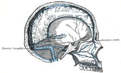 Sagittal section of the skull, showing the sinuses of the dura. | |
| Details | |
| Identifiers | |
| Latin | Sinus durae matris |
| MeSH | D003392 |
| TA | A12.3.05.101 |
| FMA | 76590 |
| Anatomical terminology | |
The dural venous sinuses (also called dural sinuses, cerebral sinuses, or cranial sinuses) are venous channels found between the endosteal and meningeal layers of dura mater in the brain.[1] They receive blood from internal and external veins of the brain, receive cerebrospinal fluid (CSF) from the subarachnoid space via arachnoid granulations, and mainly empty into the internal jugular vein.
Venous sinuses
| Name | Drains to |
| Anterior | |
| Sphenoparietal sinuses | Cavernous sinuses |
| Cavernous sinuses | Superior and inferior petrosal sinuses |
| Midline | |
| Superior sagittal sinus | Typically becomes right transverse sinus or confluence of sinuses |
| Inferior sagittal sinus | Straight sinus |
| Straight sinus | Typically becomes left transverse sinus or confluence of sinuses |
| Posterior | |
| Occipital sinus | Confluence of sinuses |
| Confluence of sinuses | Right and Left transverse sinuses |
| Lateral | |
| Superior petrosal sinus | Transverse sinuses |
| Transverse sinuses | Sigmoid sinus |
| Inferior petrosal sinus | Internal jugular vein |
| Sigmoid sinuses | Internal jugular vein |
Structure
The walls of the dural venous sinuses are composed of dura mater lined with endothelium, a specialized layer of flattened cells found in blood vessels. They differ from other blood vessels in that they lack a full set of vessel layers (e.g. tunica media) characteristic of arteries and veins. It also lacks valves as seen in arteries.
Clinical relevance
The sinuses can be injured by trauma in which damage to the dura mater, may result in blood clot formation (thrombosis) within the dural sinuses. Other common causes of dural sinus thrombosis include tracking of infection through the ophthalmic vein in orbital cellulitis. While rare, dural sinus thrombosis may lead to hemorrhagic infarction or cerebral oedema with serious consequences including epilepsy, neurological deficits, or death.[2]
Additional images
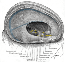 Dura mater and its processes exposed by removing part of the right half of the skull, and the brain.
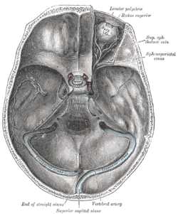 The sinuses at the base of the skull.
References
- ↑ Kiernan, John A. (2005). Barr's The Human Nervous System: An Anatomical Viewpoint. Lippincott Williams & Wilkins. pp. 428–230. ISBN 0-7817-5154-3.
- ↑ de Bruijn SF, Stam J (1999). "Randomized, placebo-controlled trial of anticoagulant treatment with low-molecular-weight heparin for cerebral sinus thrombosis". Stroke. 30 (3): 484–8. doi:10.1161/01.str.30.3.484. PMID 10066840.
External links
- http://neuroangio.org/venous-brain-anatomy/venous-sinuses/
- http://rad.usuhs.edu/medpix/parent.php3?mode=TFcase_thumbnails&pt_id=13693&quiz=no#top