Azygos vein
| Azygos vein | |
|---|---|
 Superior vena cava, inferior vena cava, azygos vein and their tributaries. (Vena azygos labeled at center.) | |
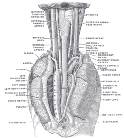 POSTERIOR VIEW: The position and relation of the esophagus in the cervical region and in the posterior mediastinum. Seen from behind. (Azygos vein labeled at bottom left.) | |
| Details | |
| Precursor | Right supracardinal vein[1] |
| Drains from | superior intercostal vein |
| Drains to | superior vena cava |
| Identifiers | |
| Latin | vena azygos |
| MeSH | D001401 |
| TA | A12.3.07.001 |
| FMA | 4838 |
| Anatomical terminology | |
The azygos vein is a vein running up the side of the thoracic vertebral column draining itself towards the superior vena cava. It connects the systems of superior vena cava and inferior vena cava and can provide an alternative path for blood to the right atrium when either of the venae cavae is blocked.[2][3]
Structure
The azygos vein transports deoxygenated blood from the posterior walls of the thorax and abdomen into the superior vena cava vein. The anatomy of this blood vessel can be quite variable. In some rare variations for example, it also drains thoracic veins, bronchial veins and even gonadal veins. The vein is so named because it has no symmetrically equivalent vein on the left side of the body.
It is formed by the union of the ascending lumbar veins with the right subcostal veins at the level of the 12th thoracic vertebra, ascending in the posterior mediastinum, and arching over the right main bronchus posteriorly at the root of the right lung to join the superior vena cava. This "arch of the azygos vein" (arcus venae azygos) is an important anatomic landmark. As an anatomical variation in 1-2% of the population, the arch can be displaced laterally, thereby creating a pleural septum separating an azygos lobe from the upper lobe of the right lung.
The anatomy of this blood vessel can be quite variable. In some rare variations for example, it also drains thoracic veins, bronchial veins and even gonadal veins. The vein is so named because it has no symmetrically equivalent vein on the left side of the body. The azygos system is considered to be the azygos vein located from rib number 2 to rib number 4 while the left part of the body has the hemiazygos vein and the accessory hemiazygos vein as the venous system.
A major tributary is the hemiazygos vein, a similar structure on the opposite side of the vertebral column. Other tributaries include the bronchial veins, pericardial veins, and posterior right intercostal veins. It communicates with the vertebrael venous plexuses.[4]
The origin and anatomical course of the azygos vein are quite variable. Usually, there is a singular azygos vein on the right side of the body. However, the azygos vein is occasionally located in the midline or two independent veins may be present like in early embryonic development.
The azygos vein generally begins at the level of the lumbar vertebrae, but may originate above the point in some case. The lumbar aspect of the azygos vein can ascend anteriorly to the lumbar vertebrae and pass behind the right crus of the diaphragm or cross the aortic hiatus (where the aorta pierces the diaphragm) to the right of the cisterna chyli, a dilated lymph sac. The common trunk of the right ascending lumbar vein and the right subcostal vein join the azygos vein anterior to the body of T12. However, if the lumbar segment is absent, this trunk may form the azygos vein.[5]

Interruption of Inferior Vena Cava (IVC) with Azygos continuation be found by frontal chest radiograph present in a soft tissue mass at the tracheobronchial angle with a smooth margin in the upper part. A tubular structure go down to the right of the spine. The aorta is located on the left. The lateral view does show a retrocardiac triangular density consistent with an IVC. Serial, contrast-enhanced CT scans of the chest and upper abdomen show an enlarged azygos vein that descends to the right of the aorta. There is no intrahepatic IVC. The liver is midline . There are multiple spleens present. [6] In developmental biology, it is proposed that azygos vein firstly drain to the posterior cardinal vein and then to longitudinal venous channel. Due to the retrogression of left common cardinal vein, the left azygos vein loses contact with posterior cardinal vein. Thus, the blood drains into the right azygos line. [7]
Function
The azygos system of veins is considered to be the azygos vein, along with its left-sided counterparts, the hemiazygos vein and the accessory hemiazygos vein.
Clinical significance
Azygos vein abnormalities can be inferenced on chest radiograph by enlargement of the azygos vein shadow greater than 1 cm. False positives can occur in heart failure causing increased pressures on the right side of the heart, or adjacent lymphadenopathy.[8] Azygos and hemiazygos continuation of the inferior vena cava (IVC) was not common in daily life. It is very hard to observe particularly when it is not associated with congenital heart disease or deep venous thrombosis. Thus, it is crucial to diagnose the enlarged azygos vein at the confluence with the superior vena cava and in the retrocrural space to prevent misdiagnosis as a right-sided paratracheal mass.The loss of the intrahepatic segment of IVC with azygous and hemiazygos continuation happens in 0.6% of patients diagnosed for congenital heart disease and usually occurs simultaneously with situs inversus, asplenia, or polysplenism, persistent left superior vena cava (SVC), and congenital pulmonary venolobar syndrome. Azygos and hemiazygos continuation of the IVC is rare especially when it is not associated with congenital heart disease.[9]
History
The Greek root zyg refers to a pair. 'A-' means not. Thus, azygos means unpaired. The azygos vein is unpaired in that there is only one in the body, mostly on the right side. While there is the hemiazygos vein and its accessory on the left side of the body, they are considered tributaries of the azygos vein rather than its left-side equivalent.
This terminology is only accurate in some species, such as the human, dog and cat. Ruminants (such as sheep and cows) have paired azygos veins.
Additional images
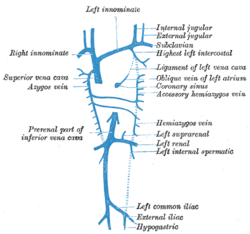 Diagram showing completion of development of the parietal veins.
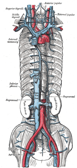 The venæ cavæ and azygos veins, with their tributaries.
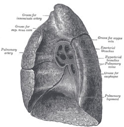 Mediastinal surface of right lung.
 Azygos vein
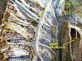 Azygos vein
See also
References
- ↑ Edward Lamperti; Michael Schuenke; Erik Schulte; Udo Schumacher; Ross, Lawrence J. (2006). General Anatomy and Musculoskeletal System (Thieme Atlas of Anatomy). Thieme Publishing Group. p. 13. ISBN 3-13-142081-2.
- ↑ Shin, Myung Soo; Ho, Kang Jey (1 July 1999). "Clinical significance of azygos vein enlargement: Radiographic recognition and etiologic analysis". Clinical Imaging. 23 (4). doi:10.1016/S0899-7071(99)00141-2.
- ↑ Piciucchi S, Barone D, Sanna S, Dubini A, Goodman LR, Oboldi D, Bertocco M, Ciccotosto C, Gavelli G, Carloni A, Poletti V (October 2014). "The azygos vein pathway: an overview from anatomical variations to pathological changes". Insights Imaging. 5 (5): 619–28. doi:10.1007/s13244-014-0351-3. PMC 4195836. PMID 25171956.
- ↑ Keith L. Moore; Arthur F. Dalley; A. M. R. Agur. Clinically Oriented Anatomy Eighth, North American Edition. Lippincott Williams & Wilkins.
- ↑ S. Standring. Gray’s Anatomy The Anatomical Basis Of Clinical Practice, 40th Edition. Elsevier Health Sciences UK.
- ↑ Van der Horst, R. L. (2015). "Interruption of the Inferior Vena Cava With Azygos Continuation". learningradiology. learningradiology. Retrieved 2018-07-09.
- ↑ "Development of superior venacava and azygous vein". slideshare. Health & Medicine, Entertainment & Humor. 2013-05-23. Retrieved 2018-06-13.
- ↑ Berman, Gerald de Lacey, Simon Morley, Laurence (2008). The chest X-ray : a survival guide. Philadelphia, PA: Saunders/Elsevier. ISBN 978-0702030468.
- ↑ Liu, Y; Guo, D; Li, J; Zhang, X; He, J; Huang, M; Dai, J; Cai, H (April 2018). "Radiological features of azygos and hemiazygos continuation of inferior vena cava: A case report". Medicine. 97 (17): e0546. doi:10.1097/MD.0000000000010546. PMC 5944491. PMID 29703035.
External links
- Anatomy figure: 19:03-03 at Human Anatomy Online, SUNY Downstate Medical Center - "Right side of the mediastinum."
- Dissection at lumc.edu
- Radiology at umich.edu
- Function at Knowyourbody.net