Thyrohyoid muscle
| Thyrohyoid muscle | |
|---|---|
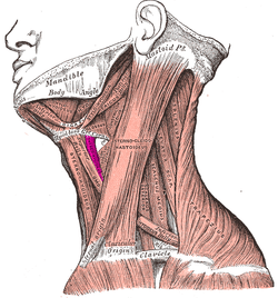 Muscles of the neck. Lateral view. (Thyrohyoideus labeled center-left.) | |
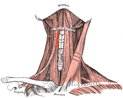 Muscles of the neck. Anterior view. (Thyrohyoideus visible center-left.) | |
| Details | |
| Origin | Thyroid cartilage of larynx |
| Insertion | Hyoid bone |
| Artery | Superior thyroid artery |
| Nerve | First cervical nerve (C1) via hypoglossal nerve |
| Actions | Elevates thyroid and depresses the hyoid bone |
| Identifiers | |
| Latin | Musculus thyrohyoideus |
| TA | A04.2.04.007 |
| FMA | 13344 |
| Anatomical terms of muscle | |
The thyrohyoid muscle is a small skeletal muscle on the neck which depresses the hyoid and elevates the larynx.
This quadrilateral muscle appearing like an upward continuation of the sternothyreoideus. It belongs to the infrahyoid muscles group.
It arises from the oblique line on the lamina of the thyroid cartilage, and is inserted into the lower border of the greater cornu of the hyoid bone.
It is innervated by the "nerve to thyrohyoid muscle". This nerve branches from the first cervical nerve as it joins the hypoglossal nerve (12th Cranial Nerve) for a short distance.
Additional images
 Hyoid bone. Anterior surface. Enlarged.
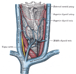 The veins of the thyroid gland.
 Hypoglossal nerve, cervical plexus, and their branches.
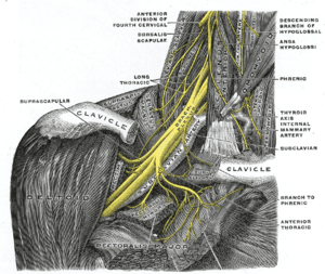 The right brachial plexus with its short branches, viewed from in front.
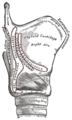 Side view of the larynx, showing muscular attachments.
 Thyrohyoid muscle
See also
References
This article incorporates text in the public domain from page 394 of the 20th edition of Gray's Anatomy (1918)
External links
- Anatomy photo:25:03-0106 at the SUNY Downstate Medical Center
- Anatomy photo:25:10-0105 at the SUNY Downstate Medical Center
- "Anatomy diagram: 25420.000-1". Roche Lexicon - illustrated navigator. Elsevier. Archived from the original on 2015-02-26.
- PTCentral
This article is issued from
Wikipedia.
The text is licensed under Creative Commons - Attribution - Sharealike.
Additional terms may apply for the media files.