Posterior triangle of the neck
| Posterior triangle of the neck | |
|---|---|
 Posterior triangle | |
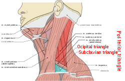
 Side of neck, showing chief surface markings. (Nerves are yellow, arteries are red.) | |
| Details | |
| Identifiers | |
| Latin |
Trigonum cervicale posterius Trigonum colli posterius Regio cervicalis posterior |
| TA | A01.2.02.009 |
| FMA | 57778 |
| Anatomical terminology | |
The posterior triangle (or lateral cervical region) is a region of the neck.
Boundaries
It has the following boundaries:
Apex: Union of the sternocleidomastoid and the trapezius muscles at the superior nuchal line of the occipital bone
Anterior: Posterior border of the sternocleidomastoideus
Posterior: Anterior border of the trapezius
Base: Middle one third of the clavicle
Roof: Investing layer of the deep cervical fascia
Floor:-(from above downward) 1.semispinalis capitis 2.spinalis capitis 3.levator scapuli 4.scleneus posterior 5.sclenues medius
Divisions
The posterior triangle is crossed, about 2.5 cm above the clavicle, by the inferior belly of the omohyoid muscle, which divides the space into two triangles:
- an upper or occipital triangle
- a lower or subclavian triangle (or supraclavicular triangle)
Contents
A) Nerves and plexuses:
- Spinal accessory nerve (Cranial Nerve XI)
- Branches of cervical plexus
- Roots and trunks of brachial plexus
- Phrenic nerve (C3,4,5)
B) Vessels:
- Subclavian artery (Third part)
- Transverse cervical artery
- Suprascapular artery
- Terminal part of external jugular vein
C) Lymph nodes:
- Occipital
- Supraclavicular
D) Muscles:
Clinical significance
The accessory nerve (CN XI) is particularly vulnerable to damage during lymph node biopsy. Damage results in an inability to shrug the shoulders or raise the arm above the head, particularly due to compromised trapezius muscle innervation.
The Internal jugular vein's superficial location within the posterior triangle also makes it vulnerable to injury.
See also
References
This article incorporates text in the public domain from page 563 of the 20th edition of Gray's Anatomy (1918)
External links
- lesson5 at The Anatomy Lesson by Wesley Norman (Georgetown University) (necktriangle)
- lesson6 at The Anatomy Lesson by Wesley Norman (Georgetown University)
- Anatomy figure: 24:01-02 at Human Anatomy Online, SUNY Downstate Medical Center - "Identification of the muscles associated with the posterolateral triangle."