Pisiform bone
| Pisiform bone | |
|---|---|
_01_palmar_view.png) Left hand anterior view (palmar view). Pisiform bone shown in red. | |
 The left pisiform bone | |
| Details | |
| Origins | ulnar collateral ligament |
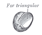
| Articulations | triangular |
| Identifiers | |
| Latin | Os pisiforme |
| MeSH | D051220 |
| TA | A02.4.08.007 |
| FMA | 23718 |
| Anatomical terms of bone | |
The pisiform bone (/ˈpaɪsɪfɔːrm/ or /ˈpɪzɪfɔːrm/), also spelled pisiforme (from the Latin pisifomis, pea-shaped), is a small knobbly, sesamoid bone that is found in the wrist. It forms the ulnar border of the carpal tunnel.
Structure
The pisiform is a sesamoid bone, with no covering membrane of periosteum. It is the last carpal bone to ossify. The pisiform bone is a small bone found in the proximal row of the wrist (carpus). It is situated where the ulna joins the wrist, within the tendon of the flexor carpi ulnaris muscle.[1]:199,205
It only has one side that acts as a joint, articulating with the triquetral bone. It is on a plane anterior to the other carpal bones and is spheroidal in form.
The pisiform bone has four surfaces:
- The dorsal surface is smooth and oval, and articulates with the triquetral: this facet approaches the superior, but not the inferior border of the bone.
- The palmar surface is rounded and rough, and gives attachment to the transverse carpal ligament, the flexor carpi ulnaris and the abductor digiti quinti.
- The lateral surface is rough, and concave.
- The medial surface' is rough and usually convex.
Function
Unlike the other carpal bones, the pisiform is not involved in movement of the wrist.[2] :5
Etymology
The etymology derives from the Latin pīsum which means "pea".
Other animals
All other tetrapods have pisiform, being the most common sesamoid.[3] As compared with apes, which have an elongated Pisiform, humans have a shorter pisiform bone. This is due to the loss of the single growth plate typically found in other mammalian pisiforms.[4] This may be because of evolutionary benefits from having a shorter pisiform bone, including facilitating ulnar deviation of the hand and preventing hyperextension of the wrist, motions that improve the action of clubbing.[5]
See also
| Wikimedia Commons has media related to Pisiform bone. |
Additional images
_-_animation01.gif) Position of pisiform bone (shown in red). Left hand. Animation.
Position of pisiform bone (shown in red). Left hand. Animation._-_animation02.gif) Pisiform bone of the left hand. Close up. Animation.
Pisiform bone of the left hand. Close up. Animation._-_animation03.gif) Pisiform bone (red) forms ulnar border of the carpal tunnel. Left hand. Animation.
Pisiform bone (red) forms ulnar border of the carpal tunnel. Left hand. Animation. Pisiform bone.
References
- ↑ Tim D. White, Human Osteology, 2nd edition (San Diego: Academic Press, 2000)
- ↑ Beasley's Surgery of the Hand. Thieme New York. 2003. ISBN 9781282950023.
- ↑ Liem (2005) functional anatomy of the vertebrates
- ↑ Kjosness, Kelsey M.; Hines, Jasmine E.; Lovejoy, C. Owen; Reno, Philip L. (November 2014). "The pisiform growth plate is lost in humans and supports a role for in growth plate formation". Journal of Anatomy. 225 (5): 527–538. doi:10.1111/joa.12235. PMC 4292754. PMID 25279687.
- ↑ Young, Richard W. (January 2003). "Evolution of the human hand: the role of throwing and clubbing". Journal of Anatomy. 202 (1): 165–174. doi:10.1046/j.1469-7580.2003.00144.x. PMC 1571064. PMID 12587931.
External links
- Cross section image: limbs/hand/hand-fr-1—Plastination Laboratory at the Medical University of Vienna
- Hand kinesiology at the University of Kansas Medical Center
- Illustration at ntu.edu.tw