Pericardiacophrenic artery
| Pericardiacophrenic artery | |
|---|---|
 The phrenic nerve and its relations with the vagus nerve. (Pericardiacophrenic artery not labeled, but region is visible.) | |
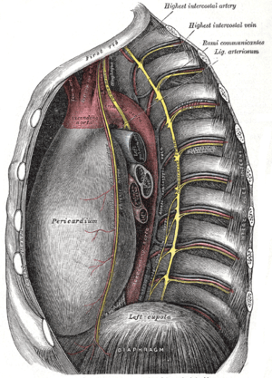 The thoracic aorta, viewed from the left side. (Pericardiacophrenic labeled at center left.) | |
| Details | |
| Source | Internal thoracic |
| Vein | pericardiacophrenic veins |
| Supplies | pericardium, thoracic diaphragm |
| Identifiers | |
| Latin | arteria pericardiacophrenica |
| TA | A12.2.08.034 |
| FMA | 3964 |
|
Anatomical terminology | |
The pericardiacophrenic artery is a long slender branch of the internal thoracic artery. It accompanies the phrenic nerve, between the pleura and pericardium, to the diaphragm, to which it is distributed. It anastomoses with the musculophrenic and superior phrenic arteries.
On their course through the thoracic cavity, the pericardiacophrenic arteries are located within and supply the fibrous pericardium.[1] Along with the musculophrenic arteries, they also provide arterial supply to the diaphragm.[2]
References
This article incorporates text in the public domain from page 584 of the 20th edition of Gray's Anatomy (1918)
External links
- Anatomy photo:19:11-0104 at the SUNY Downstate Medical Center - "Pleural Cavities and Lungs: Structures Beneath the Left Mediastinal pleura"
This article is issued from
Wikipedia.
The text is licensed under Creative Commons - Attribution - Sharealike.
Additional terms may apply for the media files.