Nanoparticle
Nanoparticles are particles between 1 and 100 nanometres (nm) in size with a surrounding interfacial layer. The interfacial layer is an integral part of nanoscale matter, fundamentally affecting all of its properties. The interfacial layer typically consists of ions, inorganic and organic molecules. Organic molecules coating inorganic nanoparticles are known as stabilizers, capping and surface ligands, or passivating agents.[1] In nanotechnology, a particle is defined as a small object that behaves as a whole unit with respect to its transport and properties. Particles are further classified according to diameter.[2]
Definition
Note 1: Modified from definitions of nanoparticle and nanogel in [refs.,[3][4]].
Note 2: The basis of the 100-nm limit is the fact that novel properties that
differentiate particles from the bulk material typically develop at a critical
length scale of under 100 nm.
Note 3: Because other phenomena (transparency or turbidity, ultrafiltration,
stable dispersion, etc.) that extend the upper limit are occasionally considered,
the use of the prefix nano is accepted for dimensions smaller than 500 nm,
provided reference to the definition is indicated.
nanoparticles.[5]
The term "nanoparticle" is not usually applied to individual molecules; it usually refers to inorganic materials.
Ultrafine particles are the same as nanoparticles and between 1 and 100 nm in size, as opposed to fine particles are sized between 100 and 2,500 nm, and coarse particles cover a range between 2,500 and 10,000 nm. The reason for the synonymous definition of nanoparticles and ultrafine particles is that, during the 1970s and 80s, when the first thorough fundamental studies with "nanoparticles" were underway in the USA (by Granqvist and Buhrman)[6] and Japan, (within an ERATO Project)[7] they were called "ultrafine particles" (UFP). However, during the 1990s before the National Nanotechnology Initiative was launched in the USA, the new name, "nanoparticle," had become more common (for example, see the same senior author's paper 20 years later addressing the same issue, lognormal distribution of sizes[8]). Nanoparticles can exhibit size-related properties significantly different from those of either fine particles or bulk materials.[9][10][11]
Nanoclusters have at least one dimension between 1 and 10 nanometers and a narrow size distribution. Nanopowders[12] are agglomerates of ultrafine particles, nanoparticles, or nanoclusters. Nanometer-sized single crystals, or single-domain ultrafine particles, are often referred to as nanocrystals.
According to ISO Technical Specification 80004, a nanoparticle is defined as a nano-object with all three external dimensions in the nanoscale, whose longest and shortest axes do not differ significantly, with a significant difference typically being a factor of at least 3.[13]
The terms colloid and nanoparticle are not interchangeable. A colloid is a mixture which has solid particles dispersed in a liquid medium. The term applies only if the particles are larger than atomic dimensions but small enough to exhibit Brownian motion, with the critical size range (or particle diameter) typically ranging from nanometers (10−9 m) to micrometers (10−6 m).[14] Colloids can contain particles too large to be nanoparticles, and nanoparticles can exist in non-colloidal form, for examples as a powder or in a solid matrix.
History
Although nanoparticles are associated with modern science, they have a long history. Nanoparticles were used by artisans as far back as Rome in the fourth century in the famous Lycurgus cup made of dichroic glass as well as the ninth century in Mesopotamia for creating a glittering effect on the surface of pots.[15][16][17] In modern times, pottery from the Middle Ages and Renaissance often retains a distinct gold- or copper-colored metallic glitter. This luster is caused by a metallic film that was applied to the transparent surface of a glazing. The luster can still be visible if the film has resisted atmospheric oxidation and other weathering.[16][17]
The luster originates within the film itself, which contains silver and copper nanoparticles dispersed homogeneously in the glassy matrix of the ceramic glaze. These nanoparticles are created by the artisans by adding copper and silver salts and oxides together with vinegar, ochre, and clay on the surface of previously-glazed pottery. The object is then placed into a kiln and heated to about 600 °C in a reducing atmosphere.[17] In heat the glaze softens, causing the copper and silver ions to migrate into the outer layers of the glaze. There the reducing atmosphere reduced the ions back to metals, which then came together forming the nanoparticles that give the color and optical effects.[17] Luster technique showed that ancient craftsmen had a sophisticated empirical knowledge of materials. The technique originated in the Muslim world. As Muslims were not allowed to use gold in artistic representations, they sought a way to create a similar effect without using real gold. The solution they found was using luster.[17][18]
Michael Faraday provided the first description, in scientific terms, of the optical properties of nanometer-scale metals in his classic 1857 paper. In a subsequent paper, the author (Turner) points out that: "It is well known that when thin leaves of gold or silver are mounted upon glass and heated to a temperature that is well below a red heat (~500 °C), a remarkable change of properties takes place, whereby the continuity of the metallic film is destroyed. The result is that white light is now freely transmitted, reflection is correspondingly diminished, while the electrical resistivity is enormously increased."[19][20][21]
Properties
Nanoparticles are of great scientific interest as they are, in effect, a bridge between bulk materials and atomic or molecular structures. A bulk material should have constant physical properties regardless of its size, but at the nano-scale size-dependent properties are often observed. Thus, the properties of materials change as their size approaches the nanoscale and as the percentage of the surface in relation to the percentage of the volume of a material becomes significant. For bulk materials larger than one micrometer (or micron), the percentage of the surface is insignificant in relation to the volume in the bulk of the material. The interesting and sometimes unexpected properties of nanoparticles are therefore largely due to the large surface area of the material, which dominates the contributions made by the small bulk of the material.
Nanoparticles often possess unexpected optical properties as they are small enough to confine their electrons and produce quantum effects.[22] For example, gold nanoparticles appear deep-red to black in solution. Nanoparticles of yellow gold and grey silicon are red in color. Gold nanoparticles melt at much lower temperatures (~300 °C for 2.5 nm size) than the gold slabs (1064 °C);.[23] Absorption of solar radiation is much higher in materials composed of nanoparticles than it is in thin films of continuous sheets of material. In both solar PV and solar thermal applications, controlling the size, shape, and material of the particles, it is possible to control solar absorption.[24][25][26][27] Recently, the core (metal)-shell (dielectric) nanoparticle has demonstrated a zero backward scattering with enhanced forward scattering on Si substrate when surface plasmon is located in front of a solar cell.[28] The core-shell nanoparticles can support simultaneously both electric and magnetic resonances, demonstrating entirely new properties when compared with bare metallic nanoparticles if the resonances are properly engineered.
Other size-dependent property changes include quantum confinement in semiconductor particles, surface plasmon resonance[22] in some metal particles and superparamagnetism in magnetic materials. What would appear ironic is that the changes in physical properties are not always desirable. Ferromagnetic materials smaller than 10 nm can switch their magnetisation direction using room temperature thermal energy, thus making them unsuitable for memory storage.[29]
Suspensions of nanoparticles are possible since the interaction of the particle surface with the solvent is strong enough to overcome density differences, which otherwise usually result in a material either sinking or floating in a liquid.
The high surface area to volume ratio of nanoparticles provides a tremendous driving force for diffusion, especially at elevated temperatures. Sintering can take place at lower temperatures, over shorter time scales than for larger particles. In theory, this does not affect the density of the final product, though flow difficulties and the tendency of nanoparticles to agglomerate complicates matters. Moreover, nanoparticles have been found to impart some extra properties to various day to day products. For example, the presence of titanium dioxide nanoparticles imparts what we call the self-cleaning effect, and, the size being nano-range, the particles cannot be observed. Zinc oxide particles have been found to have superior UV blocking properties compared to its bulk substitute. This is one of the reasons why it is often used in the preparation of sunscreen lotions,[30] is completely photostable[31] and toxic.[32] [33] [34] [35] [36] [37]
Clay nanoparticles when incorporated into polymer matrices increase reinforcement, leading to stronger plastics, verifiable by a higher glass transition temperature and other mechanical property tests. These nanoparticles are hard, and impart their properties to the polymer (plastic). Nanoparticles have also been attached to textile fibers in order to create smart and functional clothing.[38]
Metal, dielectric, and semiconductor nanoparticles have been formed, as well as hybrid structures (e.g., core–shell nanoparticles).[39] Nanoparticles made of semiconducting material may also be labeled quantum dots if they are small enough (typically sub 10 nm) that quantization of electronic energy levels occurs. Such nanoscale particles are used in biomedical applications as drug carriers or imaging agents with work being done to try to understand the fluid dynamic properties (e.g. drag forces) in nanoscale applications.[40][41] This has shown the relationship between the fluid forces on nanoparticles and the fluid Reynolds and Knudsen numbers.
_with_complete_passivation.png)
Semi-solid and soft nanoparticles have been manufactured. A prototype nanoparticle of semi-solid nature is the liposome. Various types of liposome nanoparticles are currently used clinically as delivery systems for anticancer drugs and vaccines.
Nanoparticles with one half hydrophilic and the other half hydrophobic are termed Janus particles and are particularly effective for stabilizing emulsions. They can self-assemble at water/oil interfaces and act as solid surfactants.
Hydrogel nanoparticles made of N-isopropylacrylamide hydrogel core shell can be dyed with affinity baits, internally.[42] These affinity baits allow the nanoparticles to isolate and remove undesirable proteins while enhancing the target analytes.[42]
Variation in properties
The chemical processing and synthesis of high-performance technological components for the private, industrial, and military sectors requires the use of high-purity ceramics (oxide ceramics, such as aluminium oxide or copper(II) oxide), polymers, glass-ceramics, and composite materials, as metal carbides (SiC), nitrides (Aluminum nitrides, Silicon nitride), metals (Al, Cu), non-metals (graphite, carbon nanotubes) and layered (Al + Aluminium carbonate, Cu + C). In condensed bodies formed from fine powders, the irregular particle sizes and shapes in a typical powder often lead to non-uniform packing morphologies that result in packing density variations in the powder compact.
Uncontrolled agglomeration of powders due to attractive van der Waals forces can also give rise to microstructural heterogeneity. Differential stresses that develop as a result of non-uniform drying shrinkage are directly related to the rate at which the solvent can be removed, and thus highly dependent upon the distribution of porosity. Such stresses have been associated with a plastic-to-brittle transition in consolidated bodies, and can yield to crack propagation in the unfired body if not relieved.[43][44][45]
In addition, any fluctuations in packing density in the compact as it is prepared for the kiln are often amplified during the sintering process, yielding inhomogeneous densification. Some pores and other structural defects associated with density variations have been shown to play a detrimental role in the sintering process by growing and thus limiting end-point densities. Differential stresses arising from inhomogeneous densification have also been shown to result in the propagation of internal cracks, thus becoming the strength-controlling flaws.[46][47][48]
Inert gas evaporation and inert gas deposition[6][7] are free many of these defects due to the distillation (cf. purification) nature of the process and having enough time to form single crystal particles, however even their non-aggreated deposits have lognormal size distribution, which is typical with nanoparticles.[7] The reason why modern gas evaporation techniques can produce a relatively narrow size distribution is that aggregation can be avoided.[7] However, even in this case, random residence times in the growth zone, due to the combination of drift and diffusion, result in a size distribution appearing lognormal.[8]
It would, therefore, appear desirable to process a material in such a way that it is physically uniform with regard to the distribution of components and porosity, rather than using particle size distributions that will maximize the green density. The containment of a uniformly dispersed assembly of strongly interacting particles in suspension requires total control over interparticle forces. Monodisperse nanoparticles and colloids provide this potential. [49]
Monodisperse powders of colloidal silica, for example, may therefore be stabilized sufficiently to ensure a high degree of order in the colloidal crystal or polycrystalline colloidal solid that results from aggregation. The degree of order appears to be limited by the time and space allowed for longer-range correlations to be established. Such defective polycrystalline colloidal structures would appear to be the basic elements of submicrometer colloidal materials science and, therefore, provide the first step in developing a more rigorous understanding of the mechanisms involved in microstructural evolution in high performance materials and components. [50][51]
Synthesis
There are several methods for creating nanoparticles, including gas condensation, attrition, chemical precipitation, ion implantation, pyrolysis and hydrothermal synthesis. In attrition, macro- or micro-scale particles are ground in a ball mill, a planetary ball mill, or other size-reducing mechanism. The resulting particles are air classified to recover nanoparticles. In pyrolysis, a vaporous precursor (liquid or gas) is forced through an orifice at high pressure and burned. The resulting solid (a version of soot) is air classified to recover oxide particles from by-product gases. Traditional pyrolysis often results in aggregates and agglomerates rather than single primary particles. Ultrasonic nozzle spray pyrolysis (USP) on the other hand aids in preventing agglomerates from forming.
A thermal plasma can deliver the energy to vaporize small micrometer-size particles. The thermal plasma temperatures are in the order of 10,000 K, so that solid powder easily evaporates. Nanoparticles are formed upon cooling while exiting the plasma region. The main types of the thermal plasma torches used to produce nanoparticles are dc plasma jet, dc arc plasma, and radio frequency (RF) induction plasmas. In the arc plasma reactors, the energy necessary for evaporation and reaction is provided by an electric arc formed between the anode and the cathode. For example, silica sand can be vaporized with an arc plasma at atmospheric pressure, or thin aluminum wires can be vaporized by exploding wire method. The resulting mixture of plasma gas and silica vapour can be rapidly cooled by quenching with oxygen, thus ensuring the quality of the fumed silica produced.
In RF induction plasma torches, energy coupling to the plasma is accomplished through the electromagnetic field generated by the induction coil. The plasma gas does not come in contact with electrodes, thus eliminating possible sources of contamination and allowing the operation of such plasma torches with a wide range of gases including inert, reducing, oxidizing, and other corrosive atmospheres. The working frequency is typically between 200 kHz and 40 MHz. Laboratory units run at power levels in the order of 30–50 kW, whereas the large-scale industrial units have been tested at power levels up to 1 MW. As the residence time of the injected feed droplets in the plasma is very short, it is important that the droplet sizes are small enough in order to obtain complete evaporation. The RF plasma method has been used to synthesize different nanoparticle materials, for example synthesis of various ceramic nanoparticles such as oxides, carbours/carbides, and nitrides of Ti and Si (see Induction plasma technology).
Inert-gas condensation is frequently used to make nanoparticles from metals with low melting points. The metal is vaporized in a vacuum chamber and then supercooled with an inert gas stream. The supercooled metal vapor condenses into nanometer-size particles, which can be entrained in the inert gas stream and deposited on a substrate or studied in situ.
Nanoparticles can also be formed using radiation chemistry. Radiolysis from gamma rays can create strongly active free radicals in solution. This relatively simple technique uses a minimum number of chemicals. These including water, a soluble metallic salt, a radical scavenger (often a secondary alcohol), and a surfactant (organic capping agent). High gamma doses on the order of 104 Gray are required. In this process, reducing radicals will drop metallic ions down to the zero-valence state. A scavenger chemical will preferentially interact with oxidizing radicals to prevent the re-oxidation of the metal. Once in the zero-valence state, metal atoms begin to coalesce into particles. A chemical surfactant surrounds the particle during formation and regulates its growth. In sufficient concentrations, the surfactant molecules stay attached to the particle. This prevents it from dissociating or forming clusters with other particles. Formation of nanoparticles using the radiolysis method allows for tailoring of particle size and shape by adjusting precursor concentrations and gamma dose.[52]
Sol–gel
The sol–gel process is a wet-chemical technique (also known as chemical solution deposition) widely used recently in the fields of materials science and ceramic engineering. Such methods are used primarily for the fabrication of materials (typically a metal oxide) starting from a chemical solution (sol, short for solution), which acts as the precursor for an integrated network (or gel) of either discrete particles or network polymers. [53]
Typical precursors are metal alkoxides and metal chlorides, which undergo hydrolysis and polycondensation reactions to form either a network "elastic solid" or a colloidal suspension (or dispersion) – a system composed of discrete (often amorphous) submicrometer particles dispersed to various degrees in a host fluid. Formation of a metal oxide involves connecting the metal centers with oxo (M-O-M) or hydroxo (M-OH-M) bridges, therefore generating metal-oxo or metal-hydroxo polymers in solution. Thus, the sol evolves toward the formation of a gel-like diphasic system containing both a liquid phase and solid phase whose morphologies range from discrete particles to continuous polymer networks.[54]
In the case of the colloid, the volume fraction of particles (or particle density) may be so low that a significant amount of fluid may need to be removed initially for the gel-like properties to be recognized. This can be accomplished in a number of ways. The most simple method is to allow time for sedimentation to occur, and then pour off the remaining liquid. Centrifugation can also be used to accelerate the process of phase separation.
Removal of the remaining liquid (solvent) phase requires a drying process, which typically causes shrinkage and densification. The rate at which the solvent can be removed is ultimately determined by the distribution of porosity in the gel. The ultimate microstructure of the final component will clearly be strongly influenced by changes implemented during this phase of processing. Afterward, a thermal treatment, or firing process, is often necessary in order to favor further polycondensation and enhance mechanical properties and structural stability via final sintering, densification, and grain growth. One of the distinct advantages of using this methodology as opposed to the more traditional processing techniques is that densification is often achieved at a much lower temperature.
The precursor sol can be either deposited on a substrate to form a film (e.g., by dip-coating or spin-coating), cast into a suitable container with the desired shape (e.g., to obtain a monolithic ceramics, glasses, fibers, membranes, aerogels), or used to synthesize powders (e.g., microspheres, nanospheres). The sol–gel approach is a cheap and low-temperature technique that allows for the fine control of the product’s chemical composition. Even small quantities of dopants, such as organic dyes and rare earth metals, can be introduced in the sol and end up uniformly dispersed in the final product. It can be used in ceramics processing and manufacturing as an investment casting material, or as a means of producing very thin films of metal oxides for various purposes. Sol–gel derived materials have diverse applications in optics, electronics, energy, space, (bio)sensors, medicine (e.g., controlled drug release) and separation (e.g., chromatography) technology.[55][56]
Ion implantation
Ion implantation may be used to treat the surfaces of dielectric materials such as sapphire and silica to make composites with near-surface dispersions of metal or oxide nanoparticles. See ion implantation#Ion implantation-induced nanoparticle formation
Morphology
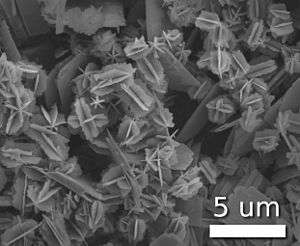
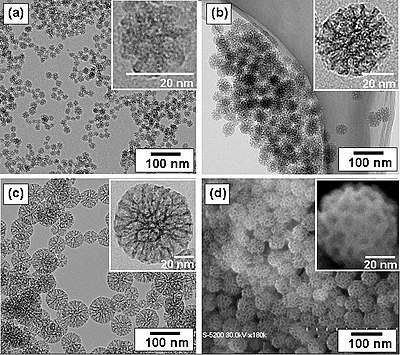
Scientists have taken to naming their particles after the real-world shapes that they might represent. Nanospheres,[57] nanochains,[58] nanoreefs,[59] nanoboxes[60] and more have appeared in the literature. These morphologies sometimes arise spontaneously as an effect of a templating or directing agent present in the synthesis such as miscellar emulsions or anodized alumina pores, or from the innate crystallographic growth patterns of the materials themselves.[61] Some of these morphologies may serve a purpose, such as long carbon nanotubes used to bridge an electrical junction, or just a scientific curiosity like the stars shown at right.
Amorphous particles usually adopt a spherical shape (due to their microstructural isotropy), whereas the shape of anisotropic microcrystalline whiskers corresponds to their particular crystal habit. At the small end of the size range, nanoparticles are often referred to as clusters. Spheres, rods, fibers, and cups are just a few of the shapes that have been grown. The study of fine particles is called micromeritics.
Characterization
Nanoparticles have different analytical requirements than conventional chemicals, for which chemical composition and concentration are sufficient metrics. Nanoparticles have other physical properties that must be measured for a complete description, such as size, shape, surface properties, crystallinity, and dispersion state. Additionally, sampling and laboratory procedures can perturb their dispersion state or bias the distribution of other properties.[62][63] In environmental contexts, an additional challenge is that many methods cannot detect low concentrations of nanoparticles that may still have an adverse effect.[62] For some applications, nanoparticles may be characterized in complex matrices such as water, soil, food, polymers, inks, complex mixtures of organic liquids such as in cosmetics, or blood.[64][65]
There are several overall categories of methods used to characterize nanoparticles. Microscopy methods generate images of individual nanoparticles to characterize their shape, size, and location. Electron microscopy and scanning probe microscopy are the dominant methods. Because nanoparticles have a size below the diffraction limit of visible light, conventional optical microscopy is not useful. Electron microscopes can be coupled to spectroscopic methods that can perform elemental analysis. Microscopy methods are destructive, and can be prone to undesirable artifacts from sample preparation, or from probe tip geometry in the case of scanning probe microscopy. Additionally, microscopy is based on single-particle measurements, meaning that large numbers of individual particles must be characterized to estimate their bulk properties.[62][64]
Spectroscopy, which measures the particles' interaction with electromagnetic radiation as a function of wavelength, is useful for some classes of nanoparticles to characterize concentration, size, and shape. X-ray, ultraviolet–visible, infrared, and nuclear magnetic resonance spectroscopy can be used with nanoparticles.[62][64] Light scattering methods using laser light, X-rays, or neutron scattering are used to determine particle size, with each method suitable for different size ranges and particle compositions.[62][64] Some miscellaneous methods are electrophoresis for surface charge, the Brunauer–Emmett–Teller method for surface area, and X-ray diffraction for crystal structure,[62] as well as mass spectrometry for particle mass, and particle counters for particle number.[64] Chromatography, centrifugation, and filtration techniques can be used to separate nanoparticles by size or other physical properties before or during characterization.[62]
Functionalization
Functionalization is the introduction of organic molecules or polymers on the surface of the nanoparticle. The surface coating of nanoparticles determines many of their physical and chemical properties, notably stability, solubility, and targeting. A coating that is multivalent or polymeric confers high stability. Functionalized nanomaterial-based catalysts can be used for catalysis of many known organic reactions.
Surface coating for biological applications
For biological applications, the surface coating should be polar to give high aqueous solubility and prevent nanoparticle aggregation. In serum or on the cell surface, highly charged coatings promote non-specific binding, whereas polyethylene glycol linked to terminal hydroxyl or methoxy groups repel non-specific interactions.[66][67] Nanoparticles can be linked to biological molecules that can act as address tags, to direct the nanoparticles to specific sites within the body,[68] specific organelles within the cell,[69] or to follow specifically the movement of individual protein or RNA molecules in living cells.[70] Common address tags are monoclonal antibodies, aptamers, streptavidin or peptides. These targeting agents should ideally be covalently linked to the nanoparticle and should be present in a controlled number per nanoparticle. Multivalent nanoparticles, bearing multiple targeting groups, can cluster receptors, which can activate cellular signaling pathways, and give stronger anchoring. Monovalent nanoparticles, bearing a single binding site,[71][72][73] avoid clustering and so are preferable for tracking the behavior of individual proteins.
Red blood cell coatings can help nanoparticles evade the immune system.[74]
Health and safety
Nanoparticles present possible dangers, both medically and environmentally.[75] [76] [77][78] Most of these are due to the high surface to volume ratio, which can make the particles very reactive or catalytic.[79] They are also able to pass through cell membranes in organisms, and their interactions with biological systems are relatively unknown.[80] However, it is unlikely the particles would enter the cell nucleus, Golgi complex, endoplasmic reticulum or other internal cellular components due to the particle size and intercellular agglomeration.[81] A recent study looking at the effects of ZnO nanoparticles on human immune cells has found varying levels of susceptibility to cytotoxicity.[82] There are concerns that pharmaceutical companies, seeking regulatory approval for nano-reformulations of existing medicines, are relying on safety data produced during clinical studies of the earlier, pre-reformulation version of the medicine. This could result in regulatory bodies, such as the FDA, missing new side effects that are specific to the nano-reformulation.[83]
Whether cosmetics and sunscreens containing nanomaterials pose health risks remains largely unknown at this stage.[84] However considerable research has demonstrated that zinc nanoparticles are not absorbed into the bloodstream in vivo.[85]
Concern has also been raised over the health effects of respirable nanoparticles from certain combustion processes.[86][87] As of 2013 the U.S. Environmental Protection Agency was investigating the safety of the following nanoparticles:[88]
- Carbon Nanotubes: Carbon materials have a wide range of uses, ranging from composites for use in vehicles and sports equipment to integrated circuits for electronic components. The interactions between nanomaterials such as carbon nanotubes and natural organic matter strongly influence both their aggregation and deposition, which strongly affects their transport, transformation, and exposure in aquatic environments. In past research, carbon nanotubes exhibited some toxicological impacts that will be evaluated in various environmental settings in current EPA chemical safety research. EPA research will provide data, models, test methods, and best practices to discover the acute health effects of carbon nanotubes and identify methods to predict them.[88]
- Cerium oxide: Nanoscale cerium oxide is used in electronics, biomedical supplies, energy, and fuel additives. Many applications of engineered cerium oxide nanoparticles naturally disperse themselves into the environment, which increases the risk of exposure. There is ongoing exposure to new diesel emissions using fuel additives containing CeO2 nanoparticles, and the environmental and public health impacts of this new technology are unknown. EPA’s chemical safety research is assessing the environmental, ecological, and health implications of nanotechnology-enabled diesel fuel additives.[88]
- Titanium dioxide: Nano titanium dioxide is currently used in many products. Depending on the type of particle, it may be found in sunscreens, cosmetics, and paints and coatings. It is also being investigated for use in removing contaminants from drinking water.[88]
- Nano Silver: Nano silver is being incorporated into textiles, clothing, food packaging, and other materials to eliminate bacteria. EPA and the U.S. Consumer Product Safety Commission are studying certain products to see whether they transfer nano-size silver particles in real-world scenarios. EPA is researching this topic to better understand how much nano-silver children come in contact with in their environments.[88]
- Iron: While nano-scale iron is being investigated for many uses, including “smart fluids” for uses such as optics polishing and as a better-absorbed iron nutrient supplement, one of its more prominent current uses is to remove contamination from groundwater. This use, supported by EPA research, is being piloted at a number of sites across the country.[88]
Applications
Scientific research on nanoparticles is intense as they have many potential applications in medicine, physics,[89][90][91] optics, and electronics.[39][25][22][26] The U.S. National Nanotechnology Initiative offers government funding focused on nanoparticle research.
The use of nanoparticles in laser dye-doped poly(methyl methacrylate) (PMMA) laser gain media was demonstrated in 2003 and it has been shown to improve conversion efficiencies and to decrease laser beam divergence.[92] Researchers attribute the reduction in beam divergence to improved dn/dT characteristics of the organic-inorganic dye-doped nanocomposite. The optimum composition reported by these researchers is 30% w/w of SiO2 (~ 12 nm) in dye-doped PMMA.
Nanoparticles are being investigated as potential drug delivery system.[93] Drugs, growth factors or other biomolecules can be conjugated to nano particles to aid targeted delivery.[94] This nanoparticle-assisted delivery allows for spatial and temporal controls of the loaded drugs to achieve the most desirable biological outcome.
Nanoparticles are also studied for possible applications as dietary supplements for delivery of biologically active substances, for example mineral elements.[95]
See also
- Ceramic engineering
- Coating
- Colloid
- Colloid-facilitated transport
- Colloidal crystal
- Colloidal gold
- Eigencolloid
- Fullerenes
- Fungal-derived nanoparticles
- Gallium(II) selenide
- Icosahedral twins
- Indium selenide
- Liposome
- Magnetic immunoassay
- Magnetic nanoparticles
- Magnetic nanochains
- Micromeritics
- Nanobiotechnology
- Nanocrystalline silicon
- Nanofluid
- Nanogeoscience
- Nanomaterials
- Nanomedicine
- Nanoparticle deposition
- Nanoparticle Tracking Analysis
- Nanotechnology
- Patchy particles
- Photonic crystal
- Plasmon
- Platinum nanoparticles
- Quantum dot
- Self-assembly of nanoparticles
- Silicon
- Silver Nano
- Sol-gel
- Transparent materials
References
- ↑ Batista, Carlos A. Silvera; Larson, Ronald G.; Kotov, Nicholas A. (2015-10-09). "Nonadditivity of nanoparticle interactions". Science. 350 (6257): 1242477. doi:10.1126/science.1242477. ISSN 0036-8075. PMID 26450215.
- ↑ Module 3: Characteristics of Particles – Particle Size Categories. epa.gov
- ↑ MacNaught, Alan D.; Wilkinson, Andrew R., eds. (1997). Compendium of Chemical Terminology: IUPAC Recommendations (2nd ed.). Blackwell Science. ISBN 978-0865426849.
- ↑ Alemán, J.; Chadwick, A. V.; He, J.; Hess, M.; Horie, K.; Jones, R. G.; Kratochvíl, P.; Meisel, I.; Mita, I.; Moad, G.; Penczek, S.; Stepto, R. F. T. (2007). "Definitions of terms relating to the structure and processing of sols, gels, networks, and inorganic-organic hybrid materials (IUPAC Recommendations 2007)". Pure and Applied Chemistry. 79 (10): 1801. doi:10.1351/pac200779101801.
- ↑ Vert, Michel; Doi, Yoshiharu; Hellwich, Karl-Heinz; Hess, Michael; Hodge, Philip; Kubisa, Przemyslaw; Rinaudo, Marguerite; Schué, François (2012). "Terminology for biorelated polymers and applications (IUPAC Recommendations 2012)" (PDF). Pure and Applied Chemistry. 84 (2): 377–410. doi:10.1351/PAC-REC-10-12-04.
- 1 2 Granqvist, C.; Buhrman, R.; Wyns, J.; Sievers, A. (1976). "Far-Infrared Absorption in Ultrafine Al Particles". Physical Review Letters. 37 (10): 625–629. Bibcode:1976PhRvL..37..625G. doi:10.1103/PhysRevLett.37.625.
- 1 2 3 4 Hayashi, C.; Uyeda, R & Tasaki, A. (1997). Ultra-fine particles: exploratory science and technology (1997 Translation of the Japan report of the related ERATO Project 1981–86). Noyes Publications.
- 1 2 Kiss, L. B.; Söderlund, J.; Niklasson, G. A.; Granqvist, C. G. (1999). "New approach to the origin of lognormal size distributions of nanoparticles". Nanotechnology. 10 (1): 25–28. Bibcode:1999Nanot..10...25K. doi:10.1088/0957-4484/10/1/006.
- ↑ Buzea, C.; Pacheco, I. I.; Robbie, K. (2007). "Nanomaterials and nanoparticles: Sources and toxicity". Biointerphases. 2 (4): MR17–MR71. doi:10.1116/1.2815690. PMID 20419892.
- ↑ ASTM E 2456 – 06 Standard Terminology Relating to Nanotechnology
- ↑ Valenti G, Rampazzo R, Bonacchi S, Petrizza L, Marcaccio M, Montalti M, Prodi L, Paolucci F (2016). "Variable Doping Induces Mechanism Swapping in Electrogenerated Chemiluminescence of Ru(bpy)32+ Core−Shell Silica Nanoparticles". J. Am. Chem. Soc. 138 (49): 15935–15942. doi:10.1021/jacs.6b08239. PMID 27960352.
- ↑ Fahlman, B. D. (2007). Materials Chemistry. Springer. pp. 282–283. ISBN 978-1-4020-6119-6.
- ↑ "ISO/TS 80004-2: Nanotechnologies — Vocabulary — Part 2: Nano-objects". International Organization for Standardization. 2015. Retrieved 2018-01-18.
- ↑ Pais, A. (2005). Subtle is the Lord: The Science and the Life of Albert Einstein. Oxford University Press. ISBN 978-0-19-280672-7. Retrieved 6 December 2016.
- ↑ "Nanotechnology Timeline | Nano". www.nano.gov. Retrieved 12 December 2016.
- 1 2 Reiss, Gunter; Hutten, Andreas (2010). "Magnetic Nanoparticles". In Sattler, Klaus D. Handbook of Nanophysics: Nanoparticles and Quantum Dots. CRC Press. pp. 2–1. ISBN 9781420075458.
- 1 2 3 4 5 Khan, Firdos Alam (2012). Biotechnology Fundamentals. CRC Press. p. 328. ISBN 9781439820094.
- ↑ Rawson, Philip S. (1984). Ceramics. University of Pennsylvania Press. ISBN 0-8122-1156-1. Retrieved 6 December 2016.
- ↑ Faraday, Michael (1857). "Experimental relations of gold (and other metals) to light". Phil. Trans. R. Soc. Lond. 147: 145–181. doi:10.1098/rstl.1857.0011.
- ↑ Beilby, G.T. (1903). "The Effects of Heat and of Solvents on Thin Films of Metal". Proceedings of the Royal Society A. 72 (477–486): 226–235. doi:10.1098/rspl.1903.0046. JSTOR 116470.
- ↑ Turner, T. (1908). "Transparent Silver and Other Metallic Films". Proceedings of the Royal Society A. 81 (548): 301–310. Bibcode:1908RSPSA..81..301T. doi:10.1098/rspa.1908.0084. JSTOR 93060.
- 1 2 3 Hewakuruppu, Y. L.; Dombrovsky, L. A.; Chen, C.; Timchenko, V.; Jiang, X.; Baek, S.; Taylor, R. A. (2013). "Plasmonic "pump–probe" method to study semi-transparent nanofluids". Applied Optics. 52 (24): 6041–6050. Bibcode:2013ApOpt..52.6041H. doi:10.1364/AO.52.006041. PMID 24085009.
- ↑ Buffat, Ph.; Borel, J.-P. (1976). "Size effect on the melting temperature of gold particles". Physical Review A. 13 (6): 2287–2298. Bibcode:1976PhRvA..13.2287B. doi:10.1103/PhysRevA.13.2287.
- ↑ Wu, Jiang; Yu, Peng; Susha, Andrei S.; Sablon, Kimberly A.; Chen, Haiyuan; Zhou, Zhihua; Li, Handong; Ji, Haining; Niu, Xiaobin (2015-04-01). "Broadband efficiency enhancement in quantum dot solar cells coupled with multispiked plasmonic nanostars". Nano Energy. 13: 827–835. doi:10.1016/j.nanoen.2015.02.012.
- 1 2 Taylor, Robert A; Otanicar, Todd; Rosengarten, Gary (2012). "Nanofluid-based optical filter optimization for PV/T systems". Light: Science & Applications. 1 (10): e34. Bibcode:2012LSA.....1E..34T. doi:10.1038/lsa.2012.34.
- 1 2 Taylor, Robert A.; Otanicar, Todd P.; Herukerrupu, Yasitha; Bremond, Fabienne; Rosengarten, Gary; Hawkes, Evatt R.; Jiang, Xuchuan; Coulombe, Sylvain (2013). "Feasibility of nanofluid-based optical filters". Applied Optics. 52 (7): 1413–22. Bibcode:2013ApOpt..52.1413T. doi:10.1364/AO.52.001413. PMID 23458793.
- ↑ Taylor, Robert A; Phelan, Patrick E; Otanicar, Todd P; Adrian, Ronald; Prasher, Ravi (2011). "Nanofluid optical property characterization: Towards efficient direct absorption solar collectors". Nanoscale Research Letters. 6 (1): 225. Bibcode:2011NRL.....6..225T. doi:10.1186/1556-276X-6-225. PMC 3211283. PMID 21711750.
- ↑ Yu, Peng; Yao, Yisen; Wu, Jiang; Niu, Xiaobin; Rogach, Andrey L.; Wang, Zhiming (2017-08-09). "Effects of Plasmonic Metal Core -Dielectric Shell Nanoparticles on the Broadband Light Absorption Enhancement in Thin Film Solar Cells". Scientific Reports. 7 (1). doi:10.1038/s41598-017-08077-9. ISSN 2045-2322.
- ↑ Gubin, Sergey P. (2009). Magnetic nanoparticles. Wiley-VCH. ISBN 3-527-40790-1.
- ↑ "Sunscreen". U.S. Food and Drug Administration. Retrieved 6 December 2016.
- ↑ Mitchnick, MA; Fairhurst, D; Pinnell, SR (1999). "Microfine zinc oxide (Z-cote) as a photostable UVA/UVB sunblock agent". Journal of the American Academy of Dermatology. 40 (1): 85–90. doi:10.1016/S0190-9622(99)70532-3. PMID 9922017.
- ↑ Heim, J; Felder, E; Tahir, MN; Kaltbeitzel, A; Heinrich, UR; Brochhausen, C; Mailänder, V; Tremel, W; Brieger, J (21 May 2015). "Genotoxic effects of zinc oxide nanoparticles". Nanoscale. 7 (19): 8931–8. Bibcode:2015Nanos...7.8931H. doi:10.1039/c5nr01167a. PMID 25916659.
- ↑ Wang, B; Zhang, Y; Mao, Z; Yu, D; Gao, C (August 2014). "Toxicity of ZnO nanoparticles to macrophages due to cell uptake and intracellular release of zinc ions". Journal of Nanoscience and Nanotechnology. 14 (8): 5688–96. doi:10.1166/jnn.2014.8876. PMID 25935990.
- ↑ Gosens, I; Kermanizadeh, A; Jacobsen, NR; Lenz, AG; Bokkers, B; de Jong, WH; Krystek, P; Tran, L; Stone, V; Wallin, H; Stoeger, T; Cassee, FR (2015). "Comparative hazard identification by a single dose lung exposure of zinc oxide and silver nanomaterials in mice". PLOS ONE. 10 (5): e0126934. doi:10.1371/journal.pone.0126934. PMC 4429007. PMID 25966284.
- ↑ Hanagata, N; Morita, H (2015). "Calcium ions rescue human lung epithelial cells from the toxicity of zinc oxide nanoparticles". The Journal of toxicological sciences. 40 (5): 625–35. doi:10.2131/jts.40.625. PMID 26354379.
- ↑ Kim, YH; Kwak, KA; Kim, TS; Seok, JH; Roh, HS; Lee, JK; Jeong, J; Meang, EH; Hong, JS; Lee, YS; Kang, JS (June 2015). "Retinopathy Induced by Zinc Oxide Nanoparticles in Rats Assessed by Micro-computed Tomography and Histopathology". Toxicological research. 31 (2): 157–63. doi:10.5487/tr.2015.31.2.157. PMC 4505346. PMID 26191382.
- ↑ Moridian, M; Khorsandi, L; Talebi, AR (2015). "Morphometric and stereological assessment of the effects of zinc oxide nanoparticles on the mouse testicular tissue". Bratislavske lekarske listy. 116 (5): 321–5. doi:10.4149/bll_2015_060. PMID 25924642.
- ↑ "The Textiles Nanotechnology Laboratory". nanotextiles.human.cornell.edu. Retrieved 6 December 2016.
- 1 2 Taylor, Robert; Coulombe, Sylvain; Otanicar, Todd; Phelan, Patrick; Gunawan, Andrey; Lv, Wei; Rosengarten, Gary; Prasher, Ravi; Tyagi, Himanshu (2013). "Small particles, big impacts: A review of the diverse applications of nanofluids". Journal of Applied Physics. 113: 011301. Bibcode:2013JAP...113a1301T. doi:10.1063/1.4754271.
- ↑ Evans, B. (January 2018). "Nano-particle drag prediction at low Reynolds number using a direct Boltzmann–BGK solution approach". Journal of Computational Physics. 352: 123–141. Bibcode:2018JCoPh.352..123E. doi:10.1016/j.jcp.2017.09.038. ISSN 0021-9991.
- ↑ Hafezi, F.; Ransing, R. S.; Lewis, R. W. (2017-02-14). "The calculation of drag on nano-cylinders". International Journal for Numerical Methods in Engineering. 111 (11): 1025–1046. Bibcode:2017IJNME.111.1025H. doi:10.1002/nme.5489. ISSN 0029-5981.
- 1 2 Luchini, A.; Geho, D.; Bishop, B.; Tran, D.; Xia, C.; Dufour, R.; et al. (2008). "Smart Hydrogel Particles: Biomarker Harvesting: One-Step Affinity Purification, Size Exclusion, and Protection against Degradation". Nano Letters. 8 (1): 350–361. Bibcode:2008NanoL...8..350L. doi:10.1021/nl072174l. PMC 2877922.
- ↑ Onoda, G.Y. Jr.; Hench, L.L., eds. (1979). Ceramic Processing Before Firing. New York: Wiley & Sons. ISBN 0-471-65410-8.
- ↑ Aksay, I.A., Lange, F.F., Davis, B.I. (1983). "Uniformity of Al2O3-ZrO2 Composites by Colloidal Filtration". J. Am. Ceram. Soc. 66 (10): C–190. doi:10.1111/j.1151-2916.1983.tb10550.x.
- ↑ Franks, G.V. & Lange, F.F. (1996). "Plastic-to-Brittle Transition of Saturated, Alumina Powder Compacts". J. Am. Ceram. Soc. 79 (12): 3161–3168. doi:10.1111/j.1151-2916.1996.tb08091.x.
- ↑ Evans, A.G. & Davidge, R.W. (1969). "The strength and fracture of fully dense polycrystalline magnesium oxide". Phil. Mag. 20 (164): 373–388. Bibcode:1969PMag...20..373E. doi:10.1080/14786436908228708.
- ↑ Evans, A. G.; Davidge, R. W. (1970). "The strength and oxidation of reaction-sintered silicon nitride". J Mat. Sci. 5 (4): 314–325. Bibcode:1970JMatS...5..314E. doi:10.1007/BF02397783.
- ↑ Lange, F.F. & Metcalf, M. (1983). "Processing-Related Fracture Origins: II, Agglomerate Motion and Cracklike Internal Surfaces Caused by Differential Sintering". J. Am. Ceram. Soc. 66 (6): 398–406. doi:10.1111/j.1151-2916.1983.tb10069.x.
- ↑ Evans, A.G. (1987). "Considerations of Inhomogeneity Effects in Sintering". J. Am. Ceram. Soc. 65 (10): 497–501. doi:10.1111/j.1151-2916.1982.tb10340.x.
- ↑ Whitesides, G.M.; et al. (1991). "Molecular Self-Assembly and Nanochemistry: A Chemical Strategy for the Synthesis of Nanostructures". Science. 254 (5036): 1312–9. Bibcode:1991Sci...254.1312W. doi:10.1126/science.1962191. PMID 1962191.
- ↑ Dabbs D. M, Aksay I.A.; Aksay (2000). "Self-Assembled Ceramics". Annu. Rev. Phys. Chem. 51: 601–22. Bibcode:2000ARPC...51..601D. doi:10.1146/annurev.physchem.51.1.601. PMID 11031294.
- ↑ Belloni, J.; Mostafavi, M.; Remita, H.; Marignier, J. L.; Delcourt, A. M. O. (1998). "Radiation-induced synthesis of mono- and multi-metallic clusters and nanocolloids". New Journal of Chemistry. 22 (11): 1239–1255. doi:10.1039/A801445K.
- ↑ Brinker, C.J. & Scherer, G.W. (1990). Sol-Gel Science: The Physics and Chemistry of Sol-Gel Processing. Academic Press. ISBN 0-12-134970-5.
- ↑ Hench, L. L.; West, J. K. (1990). "The sol-gel process". Chemical Reviews. 90: 33–72. doi:10.1021/cr00099a003.
- ↑ Klein, L. (1994). Sol-Gel Optics: Processing and Applications. Springer Verlag. ISBN 0-7923-9424-0. Retrieved 6 December 2016.
- ↑ Corriu, Robert & Anh, Nguyên Trong (2009). Molecular Chemistry of Sol-Gel Derived Nanomaterials. John Wiley and Sons. ISBN 0-470-72117-0.
- ↑ Agam, M. A.; Guo, Q (2007). "Electron Beam Modification of Polymer Nanospheres". Journal of Nanoscience and Nanotechnology. 7 (10): 3615–9. doi:10.1166/jnn.2007.814. PMID 18330181.
- ↑ Kralj, Slavko; Makovec, Darko (27 October 2015). "Magnetic Assembly of Superparamagnetic Iron Oxide Nanoparticle Clusters into Nanochains and Nanobundles". ACS Nano. 9 (10): 9700–9707. doi:10.1021/acsnano.5b02328.
- ↑ Choy J.H.; Jang E.S.; Won J.H.; Chung J.H.; Jang D.J. & Kim Y.W. (2004). "Hydrothermal route to ZnO nanocoral reefs and nanofibers". Appl. Phys. Lett. 84 (2): 287. Bibcode:2004ApPhL..84..287C. doi:10.1063/1.1639514.
- ↑ Sun, Y; Xia, Y; Xia (2002). "Shape-controlled synthesis of gold and silver nanoparticles". Science. 298 (5601): 2176–9. Bibcode:2002Sci...298.2176S. doi:10.1126/science.1077229. PMID 12481134.
- ↑ Murphy, C.J. (2002). "Materials science. Nanocubes and nanoboxes". Science. 298 (5601): 2139–41. doi:10.1126/science.1080007. PMID 12481122.
- 1 2 3 4 5 6 7 Hassellöv, Martin; Readman, James W.; Ranville, James F.; Tiede, Karen (2008-07-01). "Nanoparticle analysis and characterization methodologies in environmental risk assessment of engineered nanoparticles". Ecotoxicology. 17 (5): 344–361. doi:10.1007/s10646-008-0225-x. ISSN 0963-9292.
- ↑ Powers, Kevin W.; Palazuelos, Maria; Moudgil, Brij M.; Roberts, Stephen M. (2007-01-01). "Characterization of the size, shape, and state of dispersion of nanoparticles for toxicological studies". Nanotoxicology. 1 (1): 42–51. doi:10.1080/17435390701314902. ISSN 1743-5390.
- 1 2 3 4 5 Tiede, Karen; Boxall, Alistair B. A.; Tear, Steven P.; Lewis, John; David, Helen; Hassellöv, Martin (2008-07-01). "Detection and characterization of engineered nanoparticles in food and the environment". Food Additives & Contaminants: Part A. 25 (7): 795–821. doi:10.1080/02652030802007553. ISSN 1944-0049.
- ↑ Linsinger, Thomas P.J.; Roebben, Gert; Solans, Conxita; Ramsch, Roland. "Reference materials for measuring the size of nanoparticles". TrAC Trends in Analytical Chemistry. 30 (1): 18–27. doi:10.1016/j.trac.2010.09.005.
- ↑ Prime, KL; Whitesides, GM (1991). "Self-assembled organic monolayers: model systems for studying adsorption of proteins at surfaces". Science. 252 (5009): 1164–7. Bibcode:1991Sci...252.1164P. doi:10.1126/science.252.5009.1164. PMID 2031186.
- ↑ Liu, W; Greytak, AB; Lee, J; Wong, CR; Park, J; Marshall, LF; Jiang, W; Curtin, PN; Ting, AY; Nocera, Daniel G.; Fukumura, Dai; Jain, Rakesh K.; Bawendi, Moungi G. (2010). "Compact biocompatible quantum dots via RAFT-mediated synthesis of imidazole-based random copolymer ligand". Journal of the American Chemical Society. 132 (2): 472–83. doi:10.1021/ja908137d. PMC 2871316. PMID 20025223.
- ↑ Akerman ME, Chan WC, Laakkonen P, Bhatia SN, Ruoslahti E.; Chan; Laakkonen; Bhatia; Ruoslahti (2002). "Nanocrystal targeting in vivo". Proceedings of the National Academy of Sciences of the United States of America. 99 (20): 12617–21. Bibcode:2002PNAS...9912617A. doi:10.1073/pnas.152463399. PMC 130509. PMID 12235356.
- ↑ Hoshino, A; Fujioka, K; Oku, T; Nakamura, S; Suga, M; Yamaguchi, Y; Suzuki, K; Yasuhara, M; Yamamoto, K (2004). "Quantum dots targeted to the assigned organelle in living cells". Microbiology and immunology. 48 (12): 985–94. doi:10.1111/j.1348-0421.2004.tb03621.x. PMID 15611617.
- ↑ Suzuki, KG; Fujiwara, TK; Edidin, M; Kusumi, A (2007). "Dynamic recruitment of phospholipase Cγ at transiently immobilized GPI-anchored receptor clusters induces IP3–Ca2+ signaling: single-molecule tracking study 2". The Journal of Cell Biology. 177 (4): 731–42. doi:10.1083/jcb.200609175. PMC 2064217. PMID 17517965.
- ↑ Sung, KM; Mosley, DW; Peelle, BR; Zhang, S; Jacobson, JM (2004). "Synthesis of monofunctionalized gold nanoparticles by fmoc solid-phase reactions". Journal of the American Chemical Society. 126 (16): 5064–5. doi:10.1021/ja049578p. PMID 15099078.
- ↑ Fu, A; Micheel, CM; Cha, J; Chang, H; Yang, H; Alivisatos, AP (2004). "Discrete nanostructures of quantum dots/Au with DNA". Journal of the American Chemical Society. 126 (35): 10832–3. doi:10.1021/ja046747x. PMID 15339154.
- ↑ Howarth, M; Liu, W; Puthenveetil, S; Zheng, Y; Marshall, LF; Schmidt, MM; Wittrup, KD; Bawendi, MG; Ting, AY (2008). "Monovalent, reduced-size quantum dots for imaging receptors on living cells". Nature Methods. 5 (5): 397–9. doi:10.1038/nmeth.1206. PMC 2637151. PMID 18425138.
- ↑ "Nanoparticles play at being red blood cells". Archived from the original on 1 July 2011. Retrieved 1 July 2011.
- ↑ Zoroddu, M.A.; Medici, S.; Ledda, A.; Nurchi, V.M.; Lachowicz, J. & Peana, M. (2014). "Toxicity of nanoparticles". Curr. Med. Chem. 21 (33): 3837–53. doi:10.2174/0929867321666140601162314. PMID 25306903.
- ↑ Crisponi, G.; Nurchi, V.M.; Lachowicz, J.; Peana, M.; Medici, S.; Zoroddu, M.A. (2017). Chapter 18 - Toxicity of Nanoparticles: Etiology and Mechanisms, in Antimicrobial Nanoarchitectonics. ELSEVIER. pp. 511–546. doi:10.1016/B978-0-323-52733-0.00018-5. ISBN 9780323527330.
- ↑ Mnyusiwalla, Anisa; Daar, Abdallah S; Singer, Peter A (2003). "Mind the gap : science and ethics in nanotechnology". Nanotechnology. 14 (3): R9. Bibcode:2003Nanot..14R...9M. doi:10.1088/0957-4484/14/3/201.
- ↑ "Toxic Nanoparticles Might be Entering Human Food Supply, MU Study Finds". University of Missouri. 22 August 2013. Retrieved 23 August 2013.
- ↑ Ying, Jackie (2001). Nanostructured Materials. New York: Academic Press. ISBN 0-12-744451-3. Retrieved 6 December 2016.
- ↑ Nanotechnologies: 6. What are potential harmful effects of nanoparticles? europa.eu
- ↑ Greulich, C.; Diendorf, J.; Simon, T.; Eggeler, G.; Epple, M.; Köller, M. (2011). "Uptake and intracellular distribution of silver nanoparticles in human mesenchymal stem cells". Acta Biomaterialia. 7 (1): 347–354. doi:10.1016/j.actbio.2010.08.003. ISSN 1742-7061.
- ↑ Hanley, C; Thurber, A; Hanna, C; Punnoose, A; Zhang, J; Wingett, DG; Thurber; Hanna; Punnoose; Zhang; Wingett (2009). "The Influences of Cell Type and ZnO Nanoparticle Size on Immune Cell Cytotoxicity and Cytokine Induction". Nanoscale Res Lett. 4 (12): 1409–20. Bibcode:2009NRL.....4.1409H. doi:10.1007/s11671-009-9413-8. PMC 2894345. PMID 20652105.
- ↑ Vines T, Faunce T (2009). "Assessing the safety and cost-effectiveness of early nanodrugs". Journal of law and medicine. 16 (5): 822–45. PMID 19554862.
- ↑ Davidson, Keay (17 May 2006). "FDA urged to limit nanoparticle use in cosmetics and sunscreens". San Francisco Chronicle. Retrieved 20 April 2007.
- ↑ Benson, H., Sarveiya, V., Risk, S. and Roberts, M. S. (2005) . "Influence of anatomical site and topical formulation on skin penetration of sunscreens. Therapeutics and Clinical Risk Management, 1 3: 209–218." Retrieved 1 April 2012.
- ↑ Howard, V. (2009). "Statement of Evidence: Particulate Emissions and Health (An Bord Plenala, on Proposed Ringaskiddy Waste-to-Energy Facility)." Retrieved 26 April 2011.
- ↑ Pieters, N (March 2015). "Blood Pressure and Same-Day Exposure to Air Pollution at School: Associations with Nano-Sized to Coarse PM in Children". Environmental Health Perspectives. 123: 737–42. doi:10.1289/ehp.1408121. PMC 4492263. PMID 25756964.
- 1 2 3 4 5 6 "Nanomaterials EPA is Assessing". Environmental Protection Agency. Retrieved 6 February 2013.
- ↑ Hubler, A.; Osuagwu, O. (2010). "Digital quantum batteries: Energy and information storage in nanovacuum tube arrays". Complexity. doi:10.1002/cplx.20306.
- ↑ Stephenson, C.; Hubler, A. (2015). "Stability and conductivity of self assembled wires in a transverse electric field". Sci. Rep. 5: 15044. Bibcode:2015NatSR...515044S. doi:10.1038/srep15044.
- ↑ Hubler, A.; Lyon, D. (2013). "Gap size dependence of the dielectric strength in nano vacuum gaps". IEEE. 20: 1467–1471. doi:10.1109/TDEI.2013.6571470.
- ↑ Duarte, F. J. and James, R. O. (2003). "Tunable solid-state lasers incorporating dye-doped polymer-nanoparticle gain media". Opt. Lett. 28 (21): 2088–2090. Bibcode:2003OptL...28.2088D. doi:10.1364/OL.28.002088. PMID 14587824.
- ↑ Singh BN, Prateeksha, Gupta VK, Chen J, Atanasov AG. Organic Nanoparticle-Based Combinatory Approaches for Gene Therapy. Trends Biotechnol. 2017 Dec;35(12):1121-1124. doi: 10.1016/j.tibtech.2017.07.010. PMID 28818304.
- ↑ Wang, Zhenming; Wang, Zhefeng; Lu, William Weijia; Zhen, Wanxin; Yang, Dazhi; Peng, Songlin (2017-10-06). "Novel biomaterial strategies for controlled growth factor delivery for biomedical applications". NPG Asia Materials. 9 (10): e435. doi:10.1038/am.2017.171. ISSN 1884-4057.
- ↑ Jóźwik A, Marchewka J, Strzałkowska N, Horbańczuk JO, Szumacher-Strabel M, Cieślak A, Lipińska-Palka P, Józefiak D, Kamińska A, Atanasov AG. The Effect of Different Levels of Cu, Zn and Mn Nanoparticles in Hen Turkey Diet on the Activity of Aminopeptidases. Molecules. 2018 May 11;23(5). doi: 10.3390/molecules23051150.
Further reading
- Jackie Y. Ying (2001). Nanostructured Materials. Academic Press. pp. 5–. ISBN 978-0-12-744451-2.
- Salata, OV (2004). "Applications of nanoparticles in biology and medicine". Journal of Nanobiotechnology. 2 (1): 3. doi:10.1186/1477-3155-2-3. ISSN 1477-3155. PMC 419715. PMID 15119954.
- Nanoparticles Used In Solar Energy Conversion (ScienceDaily).
- Nanoparticles: An occupational hygiene review by RJ Aitken and others. Health and Safety Executive Research Report 274/2004
- EMERGNANO: A review of completed and near completed environment, health and safety research on nanomaterials and nanotechnology by RJ Aitken and others.
- High transmission Tandem DMA for nanoparticle studies by SEADM, 2014.
External links
| Wikibooks has a book on the topic of: Nanotechnology |
- Nanohedron.com images of nanoparticles
- Lectures on All Phases of Nanoparticle Science and Technology
- ENPRA – Risk Assessment of Engineered NanoParticles EC FP7 Project led by the Institute of Occupational Medicine