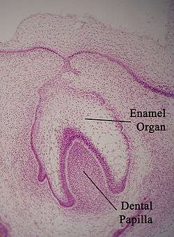
Enamel organ
| Enamel organ | |
|---|---|
 Enamel organ | |
| Details | |
| Identifiers | |
| Latin | organum enameleum |
| MeSH | D004658 |
| TE | E5.4.1.1.2.3.5 |
| Anatomical terminology | |
The enamel organ, also known as dental organ, is a cellular aggregation seen in histologic sections of a developing tooth. It lies above a condensation of ectomesenchymal cells called the dental papilla. Historically, enamel organ has been the term to describe this structure, but it was attempted unsuccessfully in recent years to change the name to dental organ in order to better represent its multiple functions apart from enamel formation.
The enamel organ functions in the formation of enamel, initiation of dentin formation, establishment of the shape of a tooth's crown, and establishment of the dentogingival junction.
The parts of the enamel organ include the inner enamel epithelium, outer enamel epithelium, stratum intermedium, and the stellate reticulum.