Dorsal aorta
| Dorsal aortae | |
|---|---|
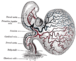 | |
 Profile view of a human embryo estimated at twenty or twenty-one days old. (Dorsal aorta labeled at center left.) | |
| Details | |
| Carnegie stage | 9 |
| Gives rise to | Descending aorta |
| System | Circulatory system |
| Identifiers | |
| Latin | aortae dorsales |
| TE | E5.11.2.1.3.0.1 |
| Anatomical terminology | |
The dorsal aortae are paired (left and right) embryological vessels which progress to form the descending aorta.[1] The paired dorsal aortae arise from aortic arches that in turn arise from the aortic sac.
Each primitive aorta anteriorly receives the vitelline vein from the yolk-sac, and is prolonged backward on the lateral aspect of the notochord under the name of the dorsal aorta.
The dorsal aortae give branches to the yolk-sac, and are continued backward through the body-stalk as the umbilical arteries to the villi of the chorion.
The two dorsal aortae combine to become the descending aorta in later development.
References
- ↑ http://www.embryology.ch/anglais/pcardio/arterien02.html. Retrieved 10 April 2017. Missing or empty
|title=(help)
External links
This article is issued from
Wikipedia.
The text is licensed under Creative Commons - Attribution - Sharealike.
Additional terms may apply for the media files.