Dental trauma
Dental trauma refers to trauma (injury) to the teeth and/or periodontium (gums, periodontal ligament, alveolar bone), and nearby soft tissues such as the lips, tongue, etc. The study of dental trauma is called dental traumatology.[1]
Prevalence
Dental trauma is most common in younger people, accounting for 17% of injuries to the body in those aged 0–6 years compared to an average of 5% across all ages.[2] It is more frequently observed in males compared to females.[3] Traumatic dental injuries are more common in permanent teeth compared to deciduous teeth and usually involve the front teeth of the upper jaw.[4]
Types
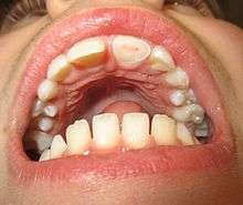
Tooth fractures[1]
- Enamel infraction
- Enamel fracture
- Enamel-dentine fracture
- Complex fracture of tooth
- Root fracture of tooth
Injuries of the periodontal apparatus
- Subluxation of the tooth (tooth knocked loose)
- Luxation of the tooth
- Intrusion of the tooth (tooth jammed into tooth socket)
- Avulsion of the tooth[5] (tooth knocked out)
Injuries to supporting bone tissues (alveolar fractures) [6][7]
This injury involves the alveolar bone and may extend beyond the alveolus. There are 5 different types of alveolar fractures:
- Communicated fracture of the socket wall
- Fracture of the socket wall
- Dentoalveolar fracture (segmental)
- Fracture of the maxilla : Le Fort fracture, zygomatic fracture, orbital blowout
- Fracture of the mandible
Trauma injuries involving the alveolus can be complicated as it does not happen in isolation, very often presents along with other types of tooth tissue injuries.
Signs of dentoalveolar fracture:
- Change to occlusion
- Multiple teeth moving together as a segment and are normally displaced
- Bruising of attached gingivae
- Gingivae across the fracture line often lacerated
Investigation : Require more than one radiographic view to identify the fracture line.
Treatment : Reposition displaced teeth under local anaesthetic and stabilise the mobile segment with a splint for 4 weeks, suture any soft tissue lacerations.
Soft tissue laceration, most commonly the lips and the gingiva.[6][7]
Soft tissues injuries are presented commonly in association with dental trauma. Areas normally affected are lips, buccal mucosa, gingivae, frenum and tongue. The most common injuries are lips and gingivae. For lips, important to rule out presence of foreign objects in wounds and lacerations through careful examination. A radiograph can be taken to identify any potential foreign objects.
Gingivae lacerations that are small normally heals spontaneously and do not require any intervention. However, this can be one of the clinical presentation of an alveolar fracture. Gingivae bleeding especially around the margins may suggest injury to the periodontal ligament of the tooth.
The facial nerve and parotid duct should be examined for any potential damage when the buccal mucosa is involved.
Deep tissue wounds should be repaired in layers with sutures that are resorbable.
Risk factors
- Young children[8]
- Sports, especially contact sports
- Piercing in tongue and lips[9]
- Military training[10][11]
- Acute changes in the barometric pressure, i.e. dental barotrauma,[12] which can affect scuba divers[13] and aviators[14]
- Class II malocclusion with increased overjet and Class II skeletal relationship [15][16]
Prevention
Regular use of a mouthguard during sports and other high-risk activities (such as military training) is the most effective prevention for dental trauma.[17] Custom made mouthguard is preferable as it fits well, provides comfort and adequate protection. However, studies in various high-risk populations for dental injuries have repeatedly reported low compliance of individuals for the regular using of mouthguard during activities.[18] Moreover, even with regular use, effectiveness of prevention of dental injuries is not complete, and injuries can still occur even when mouthguards are used as users are not always aware of the best makes or size, which inevitably result in a poor fit.[10]
One of the most important measures is to impart knowledge and awareness about dental injury to those who are involved in sports environments like boxing and in school children in which they are at high risk of suffering dental trauma through an extensive educational campaign including lectures,leaflets,Posters which should be presented in an easy understandable way.[19]
Management and future treatment options
The management depends on the type of injury involved and whether it is a baby or an adult tooth. The Dental Trauma Guide is an evidence-based and up-to-date resource to aid management of dental trauma. If teeth are completely knocked out baby front teeth should not be replaced. The area should be cleaned gently and the child brought to see a dentist. Adult front teeth (which usually erupt at around 6 years of age) can be replaced immediately if clean. See below and the Dental Trauma Guide website for more details. If a tooth is avulsed, make sure it is a permanent tooth (primary teeth should not be replanted, and instead the injury site should be cleaned to allow the adult tooth to begin to erupt).
- Reassure the patient and keep them calm.
- If the tooth can be found, pick it up by the crown (the white part). Avoid touching the root part.
- If the tooth is dirty, wash it briefly (10 seconds) under cold running water but do not scrub the tooth.
- Place the tooth back in the socket where it was lost from, taking care to place it the correct way (matching the other tooth)
- Encourage the patient to bite on a handkerchief to hold the tooth in position.
- If it is not possible to replace the tooth immediately, place it in a glass of milk or a container with the patient's saliva or in the patient's cheek (keeping it between the teeth and the inside of the cheek - note this is not suitable for young children who may swallow the tooth). Transporting the tooth in water is not recommended, as this will damage the delicate cells that make up the tooth's interior.
- Seek emergency dental treatment immediately.
The poster "Save a Tooth" is written for the public and is available in several languages—Spanish, English, Portuguese, French, Icelandic, Italian—and can be obtained at the IADT website.
When the injured teeth are painful while functioning due to damage to the periodontal ligaments (e.g., dental subluxation), a temporary splinting of the injured teeth may relieve the pain and enhance eating ability.[20] Splinting should only be used in certain situations. Splinting in lateral and extrusive luxation had a poorer prognosis than in root fractures.[21] An avulsed permanent tooth should be gently rinsed under tap water and immediately re-planted in its original socket within the alveolar bone and later temporarily splinted by a dentist.[5] Failure to re-plant the avulsed tooth within the first 40 minutes after the injury may result in very poor prognosis for the tooth.[5] Management of injured primary teeth differs from management of permanent teeth; an avulsed primary tooth should not be re-planted (to avoid damage to the permanent dental crypt).[8] This is due to the close proximity of the apex of a primary tooth to the permanent tooth underneath. The permanent dentition can suffer from tooth malformation, impacted teeth and eruption disturbances due to trauma to primary teeth. The priority should always be reducing potential damage to the underlying permanent dentition.[22]
For other injuries, it is important to keep the area clean - by using a soft toothbrush and antiseptic mouthwash such as chlorhexidine gluconate. Soft foods and avoidance of contact sports it also recommended in the short term. Dental care should be sought as quickly as possible.
Complications after management of dental trauma
Not all sequelae of trauma are immediate and many of them can occur months or years after the initial incident thus required prolonged follow-up. Common complications are pulpal necrosis, pulpal obliteration, root resorption and damage to the successors teeth in primary teeth dental trauma. The most common complication was pulp necrosis (34.2%). 50% of the tooth that have trauma related to avulsion experienced ankylotic root resorption after a median TIC (time elapsed between the traumatic event and the diagnosis of complications) of 1.18 years. Teeth that have multiple traumatic events also showed to have higher chance of pulp necrosis (61.9%) compared to teeth that experienced a single traumatic injury (25.3%) in the studies (1)[23]
Pulpal necrosis
Pulp necrosis usually occurs either as ischaemic necrosis (infarction) caused by disruption to the blood supply at the apical foramen or as an infection-related liquefactive necrosis following dental trauma (2). Signs of pulpal necrosis include[24]
- Persistent grey colour to tooth that does not fade
- Radiographic signs of periapical inflammation
- Clinical signs of infection: tenderness, sinus, suppuration, swelling
Treatment options will be extraction for the primary tooth. For the permanent tooth, endodontic treatment can be considered.
Pulpal obliteration
4-24% of traumatized teeth will have some degrees of pulpal obliteration that is characterized by the loss of pulpal space radiographically and yellow discolouration of the clinical crown. No treatment is needed if it is asymptomatic. Treatment options will be extraction for symptomatic primary tooth. For symptomatic permanent tooth, root canal treatment is often challenging due to pulp chamber is filled with calcified material and the ‘drop off’ sensation of entering a pulp chamber will not occur.[25]
Damage to the successor teeth
Dental trauma to the primary teeth might cause damage to the permanent teeth. Damage to the permanent teeth especially during development stage might have following consequences:[26]
- Crown dilaceration
- Odontoma-like malformation
- Sequestration of permanent tooth germs
- Root dilaceration
- Arrest of root formation
Primary dentition
Potential sequelae can involve pulpal necrosis, pulp obliteration and root resorption.[27] Necrosis is the most common complication and an assessment is generally made based on the colour supplemented with radiograph monitoring. A change in colour may mean that the tooth is still vital but if this persists it is likely to be non-vital.
See also
References
- 1 2 Textbook and Color Atlas of Traumatic Injuries to the Teeth, Fourth Edition, edited by Andreason J, Andreasen F, and Andersson L, Wiley-Blackwell, Oxford, UK, 2007
- ↑ Zaleckiene V, Peciuliene V, Brukiene V, Drukteinis S (2014). "Traumatic dental injuries: etiology, prevalence and possible outcomes". Stomatologija. 16 (1): 7–14. PMID 24824054.
- ↑ Kania MJ, Keeling SD, McGorray SP, Wheeler TT, King GJ (1996). "Risk factors associated with incisor injury in elementary school children". Angle Orthod. 66 (6): 423–32. doi:10.1043/0003-3219(1996)066<0423:RFAWII>2.3.CO;2. PMID 8974178.
- ↑ Granville-Garcia AF, de Menezes VA, de Lira PI (2006). "Dental trauma and associated factors in Brazilian preschoolers". Dent Traumatol. 22 (6): 318–22. doi:10.1111/j.1600-9657.2005.00390.x. PMID 17073924.
- 1 2 3 Flores MT, Andersson L, Andreasen JO, et al. (June 2007). "Guidelines for the management of traumatic dental injuries. II. Avulsion of permanent teeth". Dent Traumatol. 23 (3): 130–136. doi:10.1111/j.1600-9657.2007.00605.x. PMID 17511833.
- 1 2 Tagar, H.; Djemal, S. (September 2017). "Oral surgery II: Part 1. Acute management of dentoalveolar trauma". BDJ. 223 (6): 407–416. doi:10.1038/sj.bdj.2017.805. ISSN 1476-5373.
- 1 2 Durham, J.; Moore, U. J.; Hill, C. M.; Renton, Tara (December 2017). "Oral surgery II: Part 6. Oral and maxillofacial trauma". BDJ. 223 (12): 877–883. doi:10.1038/sj.bdj.2017.995. ISSN 1476-5373.
- 1 2 Flores MT, Malmgren B, Andersson L, et al. (August 2007). "Guidelines for the management of traumatic dental injuries. III. Primary teeth". Dent Traumatol. 23 (4): 196–202. doi:10.1111/j.1600-9657.2007.00627.x. PMID 17635351.
- ↑ Liran, Levin; Yehuda Zadik; Tal Becker (December 2005). "Oral and Dental Complications of Intra-oral Piercing". Dent Traumatol. 21 (6): 341–343. doi:10.1111/j.1600-9657.2005.00395.x. PMID 16262620.
- 1 2 Zadik Y, Levin L (December 2008). "Orofacial injuries and mouth guard use in elite commando fighters". Mil Med. 173 (12): 1185–1187. doi:10.7205/milmed.173.12.1185. PMID 19149336.
- ↑ Zadik Y, Levin L (February 2009). "Oral and facial trauma among paratroopers in the Israel Defense Forces". Dent Traumatol. 25 (1): 100–102. doi:10.1111/j.1600-9657.2008.00719.x. PMID 19208020.
- ↑ Zadik Y (Jul–Aug 2009). "Dental barotrauma". Int J Prosthodont. 22 (4): 354–7. PMID 19639071.
- ↑ Zadik, Yehuda; Drucker Scott (September 2011). "Diving dentistry: a review of the dental implications of scuba diving". Aust Dent J. 56 (3): 265–71. doi:10.1111/j.1834-7819.2011.01340.x. PMID 21884141.
- ↑ Zadik, Yehuda (January 2009). "Aviation dentistry: current concepts and practice" (PDF). British Dental Journal. 206 (1): 11–6. doi:10.1038/sj.bdj.2008.1121. PMID 19132029. Retrieved 2009-01-26.
- ↑ Borzabadi-Farahani, A.; Borzabadi-Farahani, A. (2011). "The association between orthodontic treatment need and maxillary incisor trauma, a retrospective clinical study". Oral Surgery, Oral Medicine, Oral Pathology, Oral Radiology, and Endodontology. 112 (6): e75–e80. doi:10.1016/j.tripleo.2011.05.024. PMID 21880516.
- ↑ Borzabadi-Farahani, A.; Borzabadi-Farahani, A.; Eslamipour, F. (2010). "An investigation into the association between facial profile and maxillary incisor trauma, a clinical non-radiographic study". Dental Traumatology. 26 (5): 403–408. doi:10.1111/j.1600-9657.2010.00920.x. PMID 20831636.
- ↑ Zadik Y, Levin L (February 2009). "Does a free-of-charge distribution of boil-and-bite mouthguards to young adult amateur sportsmen affect oral and facial trauma?". Dent Traumatol. 25 (1): 69–72. doi:10.1111/j.1600-9657.2008.00708.x. PMID 19208013.
- ↑ Zadik Y, Jeffet U, Levin L (December 2010). "Prevention of dental trauma in a high-risk military population: the discrepancy between knowledge and willingness to comply". Mil Med. 175 (12): 1000–1003. doi:10.7205/MILMED-D-10-00150. PMID 21265309.
- ↑ Emerich K, Nadolska-Gazda E (July 2013). "Dental trauma, prevention and knowledge concerning dental first-aid among Polish amateur boxers". J Sci Med Sport. 16 (4): 297–301. doi:10.1016/j.jsams.2012.10.002. PMID 23146163.
- ↑ Flores MT, Andersson L, Andreasen JO, et al. (April 2007). "Guidelines for the management of traumatic dental injuries. I. Fractures and luxations of permanent teeth". Dent Traumatol. 23 (2): 66–71. doi:10.1111/j.1600-9657.2007.00592.x. PMID 17367451.
- ↑ Cho, Won Chang; Nam, Ok Hyung; Kim, Mi Sun; Lee, Hyo-Seol; Choi, Sung Chul (2017-12-11). "A retrospective study of traumatic dental injuries in primary dentition: treatment outcomes of splinting". Acta Odontologica Scandinavica: 1–4. doi:10.1080/00016357.2017.1414956. ISSN 1502-3850. PMID 29228861.
- ↑ Malmgren, Barbro; Andreasen, Jens O.; Flores, Marie Therese; Robertson, Agneta; DiAngelis, Anthony J.; Andersson, Lars; Cavalleri, Giacomo; Cohenca, Nestor; Day, Peter (2017-09-15). "Guidelines for the Management of Traumatic Dental Injuries: 3. Injuries in the Primary Dentition". Pediatric Dentistry. 39 (6): 420–428. doi:10.1111/j.1600-9657.2012.01146.x. ISSN 1942-5473. PMID 29179384.
- ↑ Lin, S., Pilosof, N., Karawani, M., Wigler, R., Kaufman, A. Y., & Teich, S. T. (2016). Occurrence and timing of complications following traumatic dental injuries: A retrospective study in a dental trauma department. Journal of Clinical and Experimental Dentistry, 18(4), 429-436. [53022]. DOI: 10.4317/jced.53022
- ↑ Love RM. Effects of dental trauma on the pulp. Pract Periodontics Aesthet Dent1997;9:427–36
- ↑ McCabe, P. S. and Dummer, P. M. H. (2012), Pulp canal obliteration: an endodontic diagnosis and treatment challenge. International Endodontic Journal, 45: 177–197. doi:10.1111/j.1365-2591.2011.01963.x
- ↑ Dilaceration and Eruption Disturbances in Permanent Teeth: A Sequelae of Trauma to Their Predecessors-Diagnosis and Treatment Using Cone Beam CT.J Clin Diagn Res. 2014 May;8(5):ZD10-2. doi: 10.7860/JCDR/2014/6657.4342. Epub 2014 May 15
- ↑ Paediatric Dentistry Third Edition, edited by Richard R.Welbury, Monty S.Duggal, and Marie-Therese Hosey, Oxford, UK, 2007