Pneumatosis
Pneumatosis (pneumat- + -osis) or emphysema is the abnormal presence of air or other gas within tissues. In the lungs, emphysema involves enlargement of the distal airspaces,[1] and is a major feature of chronic obstructive pulmonary disease (COPD). Pneumoperitoneum (or peritoneal emphysema) is air or gas in the abdominal cavity, and is most commonly caused by a perforated abdominal viscus, Pneumatosis is also a frequent result of surgery.
By location
Lungs
Pulmonary emphysema
Pulmonary emphysema is a nonuniform enlargement of the distal airspaces with destruction of the acini of the alveoli, loss of lung elasticity, and (in itself) absence of any fibrotic changes.[1] It is a typical feature of chronic obstructive pulmonary disease (COPD), a type of obstructive lung disease characterized by long-term breathing problems and poor airflow.[2][3] Even without COPD, the finding of pulmonary emphysema on chest CT confers a higher mortality in tobacco smokers.[4]
Pulmonary interstitial emphysema
Pulmonary interstitial emphysema (PIE) is a collection of air outside of the normal air space of the pulmonary alveoli, found instead inside the connective tissue of the peribronchovascular sheaths, interlobular septa, and visceral pleura.
Congenital lobar emphysema
| Congenital lobar emphysema | |
|---|---|
| Classification and external resources | |
| ICD-10 | P25.0 |
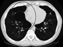
Congenital lobar emphysema (CLE), also known as congenital lobar overinflation and infantile lobar emphysema,[5] is a neonatal condition associated with enlarged air spaces in the lungs of newborn children. It is diagnosed around the time of birth or in the first 6 months of life, occurring more often in boys than girls. CLE affects the upper lung lobes more than the lower lobes, and the left lung more often than the right lung. Although CLE may be caused by abnormal development of airways (bronchi, for example) or compression of airways by nearby tissues, no cause is identified in half of cases.[6]
Other thoracic
- Pneumothorax, air or gas in the pleural space
- Pneumomediastinum, air or gas in the mediastinum
- Mediastinal emphysema, pneumomediastinum (pneumatosis/emphysema of the mediastinum)
Abdominal
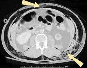
- Pneumoperitoneum (or peritoneal emphysema), air or gas in the abdominal cavity. The most common cause is a perforated abdominal viscus, generally a perforated peptic ulcer, although any part of the bowel may perforate from a benign ulcer, tumor or abdominal trauma.
- Pneumatosis intestinalis, air or gas cysts in the bowel wall
- Gastric pneumatosis, air or gas cysts in the stomach wall
Joints
Pneumarthrosis is the presence of air in a joint. Its presentation on radiography is often a vacuum cleft sign or vacuum phenomenon, which is a radiolucent cleft.
- Without disease:
Pneumarthrosis is a common normal finding in shoulders[7] as well as in sternoclavicular joints.[8] It is believed to be a cause of the sounds of joint cracking.[9] It is also a common normal postoperative finding at least in spinal surgery.[10] In fact, pneumarthrosis is extremely rare in conjunction with fluid or pus in a joint, and its presence can therefore practically exclude infection.[9]
 X-ray of a hip with prosthesis and pneumarthrosis, in this case aseptic.
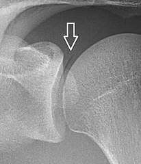 A vacuum sign is a normal finding on shoulder X-rays.
- Disease:
Pneumarthrosis is associated with osteoarthritis and spondylosis.[9]
Other locations
- Subcutaneous emphysema (gas or air trapped in the subcutaneous layer)
- Pneumarthrosis, gas in joints
References
| Look up pneumatosis in Wiktionary, the free dictionary. |
- 1 2 page 64 in: Adrian Shifren (2006). The Washington Manual Pulmonary Medicine Subspecialty Consult, Washington manual subspecialty consult series. Lippincott Williams & Wilkins. ISBN 9780781743761.
- ↑ Algusti, Alvar G.; et al. (2017). "Definition and Overview". Global Strategy for the Diagnosis, Management and Prevention of COPD. Global Initiative for Chronic Obstructive Lung Disease (GOLD). pp. 6–17.
- ↑ Roversi, Sara; Corbetta, Lorenzo; Clini, Enrico (5 May 2017). "GOLD 2017 recommendations for COPD patients: toward a more personalized approach". COPD Research and Practice. 3. doi:10.1186/s40749-017-0024-y.
- ↑ Diedtra Henderson (2014-12-16). "Emphysema on CT Without COPD Predicts Higher Mortality Risk". Medscape.
- ↑ "UpToDate: Congenital lobar emphysema". Retrieved 10 July 2016.
- ↑ Guidry, Christopher; McGahren, Eugene D. (June 2012). "Pediatric Chest I". Surgical Clinics of North America. 92 (3): 615–643. doi:10.1016/j.suc.2012.03.013. PMID 22595712.
- ↑ Dr Abhijit Datir; et al. "Vacuum phenomenon in shoulder". Radiopaedia. Retrieved 2018-01-03.
- ↑ Restrepo, Carlos S.; Martinez, Santiago; Lemos, Diego F.; Washington, Lacey; McAdams, H. Page; Vargas, Daniel; Lemos, Julio A.; Carrillo, Jorge A.; Diethelm, Lisa (2009). "Imaging Appearances of the Sternum and Sternoclavicular Joints". RadioGraphics. 29 (3): 839–859. doi:10.1148/rg.293055136. ISSN 0271-5333.
- 1 2 3 Page 60 in: Harry Griffiths (2008). Musculoskeletal Radiology. CRC Press. ISBN 9781420020663.
- ↑ Mall, J C; Kaiser, J A (1987). "The usual appearance of the postoperative lumbar spine". RadioGraphics. 7 (2): 245–269. doi:10.1148/radiographics.7.2.3448634. ISSN 0271-5333.