Hyaloid canal
| Hyaloid canal | |
|---|---|
 Horizontal section of the eyeball. (Hyaloid canal labeled running through the centre.) | |
| Details | |
| Identifiers | |
| Latin | Canalis hyaloideus |
| TA | A15.2.06.010 |
| FMA | 58837 |
| Anatomical terminology | |
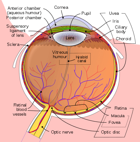
Schematic diagram of the human eye
Hyaloid canal (Cloquet's canal and Stilling's canal[1]) is a small transparent canal running through the vitreous body from the optic nerve disc to the lens. It is formed by an invagination of the hyaloid, a membrane which encloses the vitreous body.
In the fetus, the hyaloid canal contains a prolongation of the central artery of the retina, the hyaloid artery, which supplies blood to the developing lens. After birth, the hyaloid canal contains lymph and its purpose is to facilitate changes in the volume of the lens. As the lens expands in positive accommodation, its volume increases. This results in compression of the hyaloid canal, so that the volume of the eye remains constant.[2]
See also
References
- ↑ "hyaloid canal". mondofacto.com. Retrieved 20 December 2010.
- ↑ T. P. Anderson Stuart (29 March 1904). "The function of the hyaloid canal and some other new points in the mechanism of the accommodation of the eye for distance". The Journal of Physiology. 31 (1): 38–48. ISSN 0022-3751. PMC 1465472. PMID 16992721.
This article is issued from
Wikipedia.
The text is licensed under Creative Commons - Attribution - Sharealike.
Additional terms may apply for the media files.